Sunday Poster Session
Category: Colon
P0352 - Cecal Adenocarcinoma Masquerading as Perforated Appendicitis
Sunday, October 27, 2024
3:30 PM - 7:00 PM ET
Location: Exhibit Hall E

Has Audio

Samier Deen, DO
HonorHealth
Scottsdale, AZ
Presenting Author(s)
Samier Deen, DO, Faisal Mehmood, MD, Joseph Fares, MD
HonorHealth, Scottsdale, AZ
Introduction: Acute appendicitis is rarely associated with underlying malignancy.1 In the presence of concurrent cecal pathology, it is hypothesized that appendicitis is the result of luminal obstruction of the appendiceal orifice by cecal inflammation, direct tumoral occlusion, or cecal pressure due to distal colonic obstruction.1 Right hemicolectomy is appropriate in cases where possible colonic neoplasm is demonstrated on imaging, particularly in those greater than 40 years of age given an increased risk of a concomitant malignancy such as cecal adenocarcinoma.1
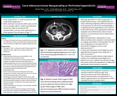
Case Description/Methods: A 45-year-old female with a past medical history of severe iron-deficiency anemia and endometriosis presented with a four-day history of right lower quadrant abdominal pain. On physical exam, she had right lower quadrant abdominal tenderness. Initial laboratory workup was remarkable for mild leukocytosis and microcytic anemia. CT abdomen and pelvis with IV contrast demonstrated findings compatible with perforated appendicitis, a fungating-appearing cecal mass measuring approximately 6.5 x 7.8 x 5.8 cm and trace pneumoperitoneum. The patient underwent laparoscopic appendectomy and hand assisted right hemicolectomy with lymphadenectomy. Tumor marker assessment included CEA which was elevated at 12.6 ng/mL along with CA 19-9, CA 27-29 and CA-125 which were within normal limits. Pathology demonstrated Stage IIIC (pT4b, pN1b, cM0) poorly differentiated invasive adenocarcinoma extending through the colonic wall and involving the terminal ileum, metastasis to two lymph nodes and negative margins. Immunohistochemical staining for microsatellite instability markers was negative. The patient was evaluated by oncology and discharged home with plan for adjuvant FOLFOX therapy in 3 weeks' time along with outpatient colonoscopy in 3 months' time.
Discussion: Although appendicitis is often recognized as a benign disease process, associated inflammation involving the cecum can make underlying malignancy difficult to discern.1 In those with coexistent cecal malignancy, prior appendectomy portends a poor prognosis in terms of survival.1 Therefore, prompt and accurate diagnostic evaluation is critical; treatment selection should consider cecal adenocarcinoma as part of the differential in older patients presenting with acute appendicitis in an effort to optimize patient survival outcomes.
References
1. Kugler et al. Appendicitis Secondary to Cecal Cancer. 2019. Pages 1-3.

Disclosures:
Samier Deen, DO, Faisal Mehmood, MD, Joseph Fares, MD. P0352 - Cecal Adenocarcinoma Masquerading as Perforated Appendicitis, ACG 2024 Annual Scientific Meeting Abstracts. Philadelphia, PA: American College of Gastroenterology.
HonorHealth, Scottsdale, AZ
Introduction: Acute appendicitis is rarely associated with underlying malignancy.1 In the presence of concurrent cecal pathology, it is hypothesized that appendicitis is the result of luminal obstruction of the appendiceal orifice by cecal inflammation, direct tumoral occlusion, or cecal pressure due to distal colonic obstruction.1 Right hemicolectomy is appropriate in cases where possible colonic neoplasm is demonstrated on imaging, particularly in those greater than 40 years of age given an increased risk of a concomitant malignancy such as cecal adenocarcinoma.1
Case Description/Methods: A 45-year-old female with a past medical history of severe iron-deficiency anemia and endometriosis presented with a four-day history of right lower quadrant abdominal pain. On physical exam, she had right lower quadrant abdominal tenderness. Initial laboratory workup was remarkable for mild leukocytosis and microcytic anemia. CT abdomen and pelvis with IV contrast demonstrated findings compatible with perforated appendicitis, a fungating-appearing cecal mass measuring approximately 6.5 x 7.8 x 5.8 cm and trace pneumoperitoneum. The patient underwent laparoscopic appendectomy and hand assisted right hemicolectomy with lymphadenectomy. Tumor marker assessment included CEA which was elevated at 12.6 ng/mL along with CA 19-9, CA 27-29 and CA-125 which were within normal limits. Pathology demonstrated Stage IIIC (pT4b, pN1b, cM0) poorly differentiated invasive adenocarcinoma extending through the colonic wall and involving the terminal ileum, metastasis to two lymph nodes and negative margins. Immunohistochemical staining for microsatellite instability markers was negative. The patient was evaluated by oncology and discharged home with plan for adjuvant FOLFOX therapy in 3 weeks' time along with outpatient colonoscopy in 3 months' time.
Discussion: Although appendicitis is often recognized as a benign disease process, associated inflammation involving the cecum can make underlying malignancy difficult to discern.1 In those with coexistent cecal malignancy, prior appendectomy portends a poor prognosis in terms of survival.1 Therefore, prompt and accurate diagnostic evaluation is critical; treatment selection should consider cecal adenocarcinoma as part of the differential in older patients presenting with acute appendicitis in an effort to optimize patient survival outcomes.
References
1. Kugler et al. Appendicitis Secondary to Cecal Cancer. 2019. Pages 1-3.

Figure: Figure A: CT abdomen and pelvis with IV contrast demonstrating fungating appearance of a cecal mass (arrow).
Figure B: Medium power field image of H&E stain demonstrating poorly differentiated invasive adenocarcinoma.
Figure C: High power field image of H&E stain demonstrating poorly differentiated invasive adenocarcinoma.
Figure B: Medium power field image of H&E stain demonstrating poorly differentiated invasive adenocarcinoma.
Figure C: High power field image of H&E stain demonstrating poorly differentiated invasive adenocarcinoma.
Disclosures:
Samier Deen indicated no relevant financial relationships.
Faisal Mehmood indicated no relevant financial relationships.
Joseph Fares indicated no relevant financial relationships.
Samier Deen, DO, Faisal Mehmood, MD, Joseph Fares, MD. P0352 - Cecal Adenocarcinoma Masquerading as Perforated Appendicitis, ACG 2024 Annual Scientific Meeting Abstracts. Philadelphia, PA: American College of Gastroenterology.
