Sunday Poster Session
Category: Colon
P0368 - Not Just Another Polyp: Rare Case of Colonic Calcifying Fibrous Tumor
Sunday, October 27, 2024
3:30 PM - 7:00 PM ET
Location: Exhibit Hall E

Has Audio
- JS
Jonathan Selzman, DO
UTHSCSA
San Antonio, TX
Presenting Author(s)
Jonathan Selzman, DO1, Jaya Vasudevan, MD2, Gabriel Magraner, MD3
1UTHSCSA, San Antonio, TX; 2University of Texas Health Science Center, San Antonio, TX; 3University of Texas Health San Antonio, San Antonio, TX
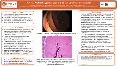
Introduction: Calcifying fibrous tumors (CFTs) are rare, benign mesenchymal neoplasms that can develop in the gastrointestinal tract and have been reported to arise in the stomach, small bowel, and colon. These uncommon lesions may present asymptomatically and are usually found as an incidental finding on endoscopy. We present a case of a 45-year-old patient diagnosed with a CFT on colonoscopy.
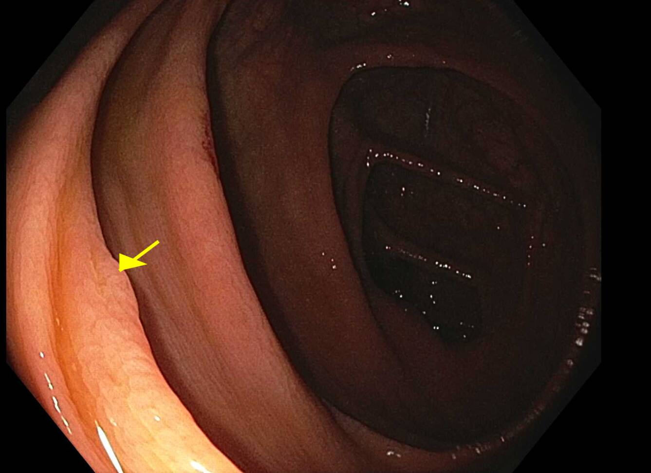
Case Description/Methods: A 45-year-old-male with past medical history of hypertension and reported irritable bowel syndrome presented to the outpatient GI clinic for an average risk screening colonoscopy. On GI review of systems, he denied abdominal pain, alteration in bowel habits, weight loss, or blood in his stool. He denied previous abdominal surgeries. On endoscopic evaluation, a 5 mm polyp in the ascending colon and a 5 mm polyp at the hepatic flexure (Figure 1) were identified and removed via cold snare polypectomy. While his ascending colon polyp microscopic studies were consistent with a hyperplastic polyp, his 5mm hepatic flexure polyp was found to be a CFT. Immunohistochemical stains utilizing S-100, CD68, Factor 13A, CD34, vimentim, SMA and CD117 supported this diagnosis. The patient was recommended to repeat colonoscopy in 5 years for surveillance.
Discussion: While rare in overall incidence, the colon is an uncommon site of CFTs, accounting for approximately 1.2% of all sites of this tumor type. The pathogenesis of CFTs is uncertain, but previous infection, trauma, or surgery, chronic tissue injury, or underlying IgG4 related disease may be risk factors. With a predilection for middle aged adults with a female predominance, CFTs are usually asymptomatic but may rarely cause symptoms such as abdominal pain, altered bowel habits, or loss of appetite via mass effect. On endoscopy, CFTs appear as well-circumscribed, unencapsulated masses with varying degrees of calcification which may hint at a preliminary diagnosis. While benign in nature with no reported malignant transformation, but identification with immunohistochemical staining is paramount to differentiate CFTs from other mesenchymal tumors, such as a sclerosing calcified gastrointestinal stromal tumors, schwannomas, leiomyomas, inflammatory myofibroblastic tumors, or solitary fibrous tumors. CFTs are cured by local resection with known recurrence in up to 20% of cases. Though no surveillance guidelines are available, for small, asymptomatic lesions, long term follow-up with repeat colonoscopy may be sufficient.

Disclosures:
Jonathan Selzman, DO1, Jaya Vasudevan, MD2, Gabriel Magraner, MD3. P0368 - Not Just Another Polyp: Rare Case of Colonic Calcifying Fibrous Tumor, ACG 2024 Annual Scientific Meeting Abstracts. Philadelphia, PA: American College of Gastroenterology.
1UTHSCSA, San Antonio, TX; 2University of Texas Health Science Center, San Antonio, TX; 3University of Texas Health San Antonio, San Antonio, TX
Introduction: Calcifying fibrous tumors (CFTs) are rare, benign mesenchymal neoplasms that can develop in the gastrointestinal tract and have been reported to arise in the stomach, small bowel, and colon. These uncommon lesions may present asymptomatically and are usually found as an incidental finding on endoscopy. We present a case of a 45-year-old patient diagnosed with a CFT on colonoscopy.
Case Description/Methods: A 45-year-old-male with past medical history of hypertension and reported irritable bowel syndrome presented to the outpatient GI clinic for an average risk screening colonoscopy. On GI review of systems, he denied abdominal pain, alteration in bowel habits, weight loss, or blood in his stool. He denied previous abdominal surgeries. On endoscopic evaluation, a 5 mm polyp in the ascending colon and a 5 mm polyp at the hepatic flexure (Figure 1) were identified and removed via cold snare polypectomy. While his ascending colon polyp microscopic studies were consistent with a hyperplastic polyp, his 5mm hepatic flexure polyp was found to be a CFT. Immunohistochemical stains utilizing S-100, CD68, Factor 13A, CD34, vimentim, SMA and CD117 supported this diagnosis. The patient was recommended to repeat colonoscopy in 5 years for surveillance.
Discussion: While rare in overall incidence, the colon is an uncommon site of CFTs, accounting for approximately 1.2% of all sites of this tumor type. The pathogenesis of CFTs is uncertain, but previous infection, trauma, or surgery, chronic tissue injury, or underlying IgG4 related disease may be risk factors. With a predilection for middle aged adults with a female predominance, CFTs are usually asymptomatic but may rarely cause symptoms such as abdominal pain, altered bowel habits, or loss of appetite via mass effect. On endoscopy, CFTs appear as well-circumscribed, unencapsulated masses with varying degrees of calcification which may hint at a preliminary diagnosis. While benign in nature with no reported malignant transformation, but identification with immunohistochemical staining is paramount to differentiate CFTs from other mesenchymal tumors, such as a sclerosing calcified gastrointestinal stromal tumors, schwannomas, leiomyomas, inflammatory myofibroblastic tumors, or solitary fibrous tumors. CFTs are cured by local resection with known recurrence in up to 20% of cases. Though no surveillance guidelines are available, for small, asymptomatic lesions, long term follow-up with repeat colonoscopy may be sufficient.

Figure: Figure 1. Colonoscopy image of 5 mm polyp at the hepatic flexure.
Disclosures:
Jonathan Selzman indicated no relevant financial relationships.
Jaya Vasudevan indicated no relevant financial relationships.
Gabriel Magraner indicated no relevant financial relationships.
Jonathan Selzman, DO1, Jaya Vasudevan, MD2, Gabriel Magraner, MD3. P0368 - Not Just Another Polyp: Rare Case of Colonic Calcifying Fibrous Tumor, ACG 2024 Annual Scientific Meeting Abstracts. Philadelphia, PA: American College of Gastroenterology.
