Sunday Poster Session
Category: Colon
P0275 - A Rare Complication: Sigmoid Diverticulitis Leading to Hepatic Abscess
Sunday, October 27, 2024
3:30 PM - 7:00 PM ET
Location: Exhibit Hall E

Has Audio
- HM
Hari Movva, MD
University of Texas Medical Branch
Galveston, TX
Presenting Author(s)
Hari Movva, MD, Giri Movva, MD, Nghia Nguyen, MD, Gurinder Luthra, MD
University of Texas Medical Branch, Galveston, TX
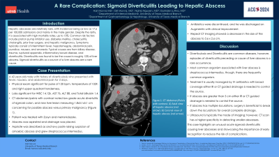
Introduction: Hepatic abscesses are relatively rare, with incidence being as low as 17.6 per 100,000 admissions, typically in males. Despite the rarity, it is associated with high mortality rates, up to 15%. Common risk factors include proton pump inhibitor use, diabetes mellitus, cholecystitis, cholangitis, prior liver surgery, and hepatic malignancy. Symptoms typically consist of intermittent fever, hepatomegaly, abdominal pain, jaundice, nausea, and anorexia. Typical causes are from biliary disease, trauma, ruptured appendix, inflammatory bowel disease, and diverticulitis. Diverticulitis was found to be the cause in roughly 12% of liver abscess with less than 20 cases being due to sigmoid diverticulitis.
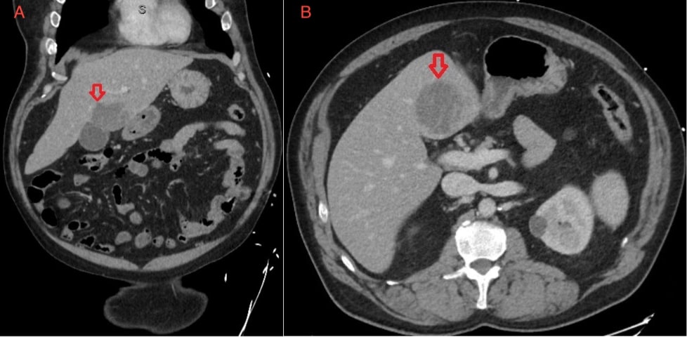
Case Description/Methods: A 60-year-old male with significant past medical history of diverticulosis who presented with fever, nausea, and abdominal pain for 3 days. On physical exam, pulse was 120 bpm, temperature of 102, and right upper quadrant tenderness. Lab data significant for leukocytosis of 14.12, AST of 76, ALT of 88, and T bili of 1.4. CT abdomen/pelvis with contrast noted low-grade acute diverticulitis of sigmoid colon, new liver lesion measuring 1.8 x 5.1 x 5.1 cm concern for possible abscess versus primary malignancy (Figure 1). Patient was treated with Zosyn and metronidazole. Abscess was aspirated and drainage was placed. Aspirate was described as anchovy paste raising suspicion of amoebic abscess and grew Streptococcus intermedius. Antibiotics were de-escalated and patient was discharged on Augmentin with clinical improvement. Repeat CT imaging showed decreased size of abscess to 3.6 x 2.6 cm.
Discussion: Diverticulosis and diverticulitis are common diseases. However, episodes of diverticulitis preceding, or cause of liver abscess is a rare occurrence. Most common organism associated with liver abscess is Streptococcus intermedius, though, there are frequently common organisms. Treatment is usually managed by IV antibiotics with broad coverage including anaerobes. If abscess is greater than 5 cm, either IR or CT guided drainage is needed to control the source. If abscess has multiple loculations, surgery is beneficial to break down the loculations for overall complete drainage. Ultrasound is typically the mode of imaging; however, CT scan has a higher specificity in detecting smaller abscesses. This case highlights an unusual acute sigmoid diverticulitis causing liver abscesses and showcasing the importance of early recognition to reduce the risk of complications.

Disclosures:
Hari Movva, MD, Giri Movva, MD, Nghia Nguyen, MD, Gurinder Luthra, MD. P0275 - A Rare Complication: Sigmoid Diverticulitis Leading to Hepatic Abscess, ACG 2024 Annual Scientific Meeting Abstracts. Philadelphia, PA: American College of Gastroenterology.
University of Texas Medical Branch, Galveston, TX
Introduction: Hepatic abscesses are relatively rare, with incidence being as low as 17.6 per 100,000 admissions, typically in males. Despite the rarity, it is associated with high mortality rates, up to 15%. Common risk factors include proton pump inhibitor use, diabetes mellitus, cholecystitis, cholangitis, prior liver surgery, and hepatic malignancy. Symptoms typically consist of intermittent fever, hepatomegaly, abdominal pain, jaundice, nausea, and anorexia. Typical causes are from biliary disease, trauma, ruptured appendix, inflammatory bowel disease, and diverticulitis. Diverticulitis was found to be the cause in roughly 12% of liver abscess with less than 20 cases being due to sigmoid diverticulitis.
Case Description/Methods: A 60-year-old male with significant past medical history of diverticulosis who presented with fever, nausea, and abdominal pain for 3 days. On physical exam, pulse was 120 bpm, temperature of 102, and right upper quadrant tenderness. Lab data significant for leukocytosis of 14.12, AST of 76, ALT of 88, and T bili of 1.4. CT abdomen/pelvis with contrast noted low-grade acute diverticulitis of sigmoid colon, new liver lesion measuring 1.8 x 5.1 x 5.1 cm concern for possible abscess versus primary malignancy (Figure 1). Patient was treated with Zosyn and metronidazole. Abscess was aspirated and drainage was placed. Aspirate was described as anchovy paste raising suspicion of amoebic abscess and grew Streptococcus intermedius. Antibiotics were de-escalated and patient was discharged on Augmentin with clinical improvement. Repeat CT imaging showed decreased size of abscess to 3.6 x 2.6 cm.
Discussion: Diverticulosis and diverticulitis are common diseases. However, episodes of diverticulitis preceding, or cause of liver abscess is a rare occurrence. Most common organism associated with liver abscess is Streptococcus intermedius, though, there are frequently common organisms. Treatment is usually managed by IV antibiotics with broad coverage including anaerobes. If abscess is greater than 5 cm, either IR or CT guided drainage is needed to control the source. If abscess has multiple loculations, surgery is beneficial to break down the loculations for overall complete drainage. Ultrasound is typically the mode of imaging; however, CT scan has a higher specificity in detecting smaller abscesses. This case highlights an unusual acute sigmoid diverticulitis causing liver abscesses and showcasing the importance of early recognition to reduce the risk of complications.

Figure: Figure 1: CT Abdomen Pelvis with contrast. A) Axial view of hepatic abscess (red arrow). B) Coronal view of hepatic abscess (red arrow).
Disclosures:
Hari Movva indicated no relevant financial relationships.
Giri Movva indicated no relevant financial relationships.
Nghia Nguyen indicated no relevant financial relationships.
Gurinder Luthra indicated no relevant financial relationships.
Hari Movva, MD, Giri Movva, MD, Nghia Nguyen, MD, Gurinder Luthra, MD. P0275 - A Rare Complication: Sigmoid Diverticulitis Leading to Hepatic Abscess, ACG 2024 Annual Scientific Meeting Abstracts. Philadelphia, PA: American College of Gastroenterology.
