Sunday Poster Session
Category: Biliary/Pancreas
P0188 - A Fortunate Case of Pyelonephritis Leads to Discovery of Rare Pancreatic Tumor
Sunday, October 27, 2024
3:30 PM - 7:00 PM ET
Location: Exhibit Hall E

Has Audio
- VD
Victoria Diaz, MD
Valley Hospital Medical Center
Las Vegas, NV
Presenting Author(s)
Victoria Diaz, MD1, Stephen Sinclair, DO2
1Valley Hospital Medical Center, Las Vegas, NV; 2Sunrise Health GME Consortium, Las Vegas, NV
Introduction: Solid pseudopapillary neoplasms (SPN) account for 1-3% of pancreatic tumors. SPN have malignant potential with metastasis in 10-15% of cases. Discovery of SPN on imaging should prompt urgent surgical evaluation.
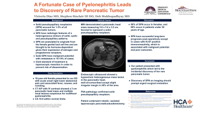
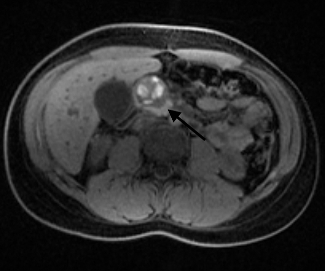
Case Description/Methods: An 18-year-old female presented to our ED with acute onset right lower abdominal pain and associated fever, nausea, and vomiting. Initial labs showed leukocytosis, with hepatic panel and lipase within normal limits. CT A/P with IV contrast showed a 3 cm pancreatic head mass and multiple renal lesions suspicious for multifocal pyelonephritis. MRI demonstrated a pancreatic head mass measuring 3.6 x 3.4 x 3.2 cm, favored to represent a solid pseudopapillary neoplasm. CA 19-9 was within normal limits. Differential diagnoses included pancreatic pseudocyst, mucinous cystic neoplasm, or intraductal papillary mucinous neoplasm. Endoscopic ultrasound showed a hypoechoic heterogeneous mass lesion in the pancreatic head, mostly well-circumscribed except slight irregular margin in 20% of the area. FNA was performed by a multi-pass technique and pathology confirmed a solid pseudopapillary neoplasm. The patient underwent robotic-assisted laparoscopic pancreaticoduodenectomy.
Discussion: Our patient presented with pyelonephritis which led to the incidental discovery of her rare pancreatic tumor. The incidence of SPN have been rising due to more frequent use of imaging modalities. SPN are postulated to originate from the female genital bud cell line and are thought to be hormone-dependent tumors given their expression of estrogen and progesterone receptors. Their predominance in females supports these hypotheses. Literature review by Vassos et al. found that 80% of SPN occurred in females, and that 85% occurred in patients under 30 years of age. SPN present with abdominal pain or compressive symptoms if large enough; however, most are discovered incidentally. SPN have radiologic features of a heterogeneous mixture of solid, cystic and pseudopapillary patterns that are well-encapsulated. Both solid and pseudopapillary proliferation are observed on histology. Tumor growth is suspected to be secondary to alterations in E-cadherin and Beta-catenin mutations which have been detected in immunohistochemistry. The gold standard of treatment is laparoscopic resection in order to prevent risk of dissemination. With the exception of Ki-67 positive immunoreactivity that predicts malignant potential and poor outcomes, most SPN have successful long-term prognosis post-operatively.

Disclosures:
Victoria Diaz, MD1, Stephen Sinclair, DO2. P0188 - A Fortunate Case of Pyelonephritis Leads to Discovery of Rare Pancreatic Tumor, ACG 2024 Annual Scientific Meeting Abstracts. Philadelphia, PA: American College of Gastroenterology.
1Valley Hospital Medical Center, Las Vegas, NV; 2Sunrise Health GME Consortium, Las Vegas, NV
Introduction: Solid pseudopapillary neoplasms (SPN) account for 1-3% of pancreatic tumors. SPN have malignant potential with metastasis in 10-15% of cases. Discovery of SPN on imaging should prompt urgent surgical evaluation.
Case Description/Methods: An 18-year-old female presented to our ED with acute onset right lower abdominal pain and associated fever, nausea, and vomiting. Initial labs showed leukocytosis, with hepatic panel and lipase within normal limits. CT A/P with IV contrast showed a 3 cm pancreatic head mass and multiple renal lesions suspicious for multifocal pyelonephritis. MRI demonstrated a pancreatic head mass measuring 3.6 x 3.4 x 3.2 cm, favored to represent a solid pseudopapillary neoplasm. CA 19-9 was within normal limits. Differential diagnoses included pancreatic pseudocyst, mucinous cystic neoplasm, or intraductal papillary mucinous neoplasm. Endoscopic ultrasound showed a hypoechoic heterogeneous mass lesion in the pancreatic head, mostly well-circumscribed except slight irregular margin in 20% of the area. FNA was performed by a multi-pass technique and pathology confirmed a solid pseudopapillary neoplasm. The patient underwent robotic-assisted laparoscopic pancreaticoduodenectomy.
Discussion: Our patient presented with pyelonephritis which led to the incidental discovery of her rare pancreatic tumor. The incidence of SPN have been rising due to more frequent use of imaging modalities. SPN are postulated to originate from the female genital bud cell line and are thought to be hormone-dependent tumors given their expression of estrogen and progesterone receptors. Their predominance in females supports these hypotheses. Literature review by Vassos et al. found that 80% of SPN occurred in females, and that 85% occurred in patients under 30 years of age. SPN present with abdominal pain or compressive symptoms if large enough; however, most are discovered incidentally. SPN have radiologic features of a heterogeneous mixture of solid, cystic and pseudopapillary patterns that are well-encapsulated. Both solid and pseudopapillary proliferation are observed on histology. Tumor growth is suspected to be secondary to alterations in E-cadherin and Beta-catenin mutations which have been detected in immunohistochemistry. The gold standard of treatment is laparoscopic resection in order to prevent risk of dissemination. With the exception of Ki-67 positive immunoreactivity that predicts malignant potential and poor outcomes, most SPN have successful long-term prognosis post-operatively.

Figure: MRI demonstrates a pancreatic head mass measuring 3.6 x 3.4 x 3.2 cm, favored to represent a solid pseudopapillary neoplasm
Disclosures:
Victoria Diaz indicated no relevant financial relationships.
Stephen Sinclair indicated no relevant financial relationships.
Victoria Diaz, MD1, Stephen Sinclair, DO2. P0188 - A Fortunate Case of Pyelonephritis Leads to Discovery of Rare Pancreatic Tumor, ACG 2024 Annual Scientific Meeting Abstracts. Philadelphia, PA: American College of Gastroenterology.
