Sunday Poster Session
Category: Biliary/Pancreas
P0198 - Recognizing Cholecystocolonic Fistula: A Rare Gallbladder Disease Complication Discovered During Routine Colonoscopy
Sunday, October 27, 2024
3:30 PM - 7:00 PM ET
Location: Exhibit Hall E

Has Audio
- HS
Hussam Sabbagh, MD
Wayne State University School of Medicine / Detroit Medical Center
Detroit, MI
Presenting Author(s)
Award: Presidential Poster Award
Hussam Sabbagh, MD1, Anshu Wadehra, MD1, Fadi Antaki, MD2, Ziad Kanaan, MD, PhD3
1Wayne State University School of Medicine / Detroit Medical Center, Detroit, MI; 2John D. Dingell VA Medical Center and Wayne State University School of Medicine, Detroit, MI; 3John D. Dingell VA Medical Center, Detroit, MI
Introduction: Cholecystoenteric fistula is a rare complication of gallbladder disease, with an incidence ranging from 0.06% to 0.14% among patients with biliary disease. This condition is most discovered intraoperatively during cholecystectomy. The duodenum is the most frequent site for these fistulas, followed by the colon. Patients with this complication have increased morbidity and mortality, making early recognition crucial. We hereby present a unique case of cholecystocolonic fistula identified during a routine outpatient colonoscopy.
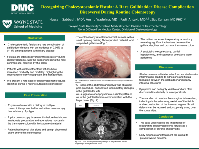
Case Description/Methods: A 71-year-old male with a history of multiple comorbidities presented for an outpatient colonoscopy due to a history of polyps. His last colonoscopy, conducted three months prior, showed inadequate preparation and edematous mucosa in the transverse colon with thick purulent material seen. On presentation, the patient's vital signs were normal. Physical examination revealed a soft, non-tender, and non-distended abdomen. Colonoscopy revealed abnormal mucosa with small opening draining fibrinopurulent material and suspected gallstones (Fig. 1). Diverticular abscess or cholecystocolonic fistula was suspected. A CT scan of the abdomen and pelvis showed inflammatory changes in the gallbladder with air, suggestive of either emphysematous cholecystitis or air in the gallbladder due to communication with the large bowel (Fig. 2). The patient was admitted and treated with antibiotics. Exploratory laparotomy was performed and revealed significant adhesions between the gallbladder, the liver and the proximal transverse colon. Sub-total cholecystectomy, partial hepatectomy, and segmental colectomy were performed. Examination of the resected specimen confirmed cholecystocolonic fistula.
Discussion: Cholecystocolonic fistulas are an uncommon complication of gallbladder disease, typically arising from pericholecystic inflammation causing adhesions between the gallbladder and intestinal structures. The clinical presentation is highly variable, often leading to discovery intraoperatively during cholecystectomy. First-line treatment is surgical, involving cholecystectomy, excision of the fistula, and reconstruction of the affected organ. Endoscopic therapy is also described, using through-the-scope clips for small defects (< 1 cm) or over-the-scope clips for defects up to 2 cm. This case illustrates an uncommon presentation of this pathology, which should not be ignored as early recognition and management are essential.

Disclosures:
Hussam Sabbagh, MD1, Anshu Wadehra, MD1, Fadi Antaki, MD2, Ziad Kanaan, MD, PhD3. P0198 - Recognizing Cholecystocolonic Fistula: A Rare Gallbladder Disease Complication Discovered During Routine Colonoscopy, ACG 2024 Annual Scientific Meeting Abstracts. Philadelphia, PA: American College of Gastroenterology.
Hussam Sabbagh, MD1, Anshu Wadehra, MD1, Fadi Antaki, MD2, Ziad Kanaan, MD, PhD3
1Wayne State University School of Medicine / Detroit Medical Center, Detroit, MI; 2John D. Dingell VA Medical Center and Wayne State University School of Medicine, Detroit, MI; 3John D. Dingell VA Medical Center, Detroit, MI
Introduction: Cholecystoenteric fistula is a rare complication of gallbladder disease, with an incidence ranging from 0.06% to 0.14% among patients with biliary disease. This condition is most discovered intraoperatively during cholecystectomy. The duodenum is the most frequent site for these fistulas, followed by the colon. Patients with this complication have increased morbidity and mortality, making early recognition crucial. We hereby present a unique case of cholecystocolonic fistula identified during a routine outpatient colonoscopy.
Case Description/Methods: A 71-year-old male with a history of multiple comorbidities presented for an outpatient colonoscopy due to a history of polyps. His last colonoscopy, conducted three months prior, showed inadequate preparation and edematous mucosa in the transverse colon with thick purulent material seen. On presentation, the patient's vital signs were normal. Physical examination revealed a soft, non-tender, and non-distended abdomen. Colonoscopy revealed abnormal mucosa with small opening draining fibrinopurulent material and suspected gallstones (Fig. 1). Diverticular abscess or cholecystocolonic fistula was suspected. A CT scan of the abdomen and pelvis showed inflammatory changes in the gallbladder with air, suggestive of either emphysematous cholecystitis or air in the gallbladder due to communication with the large bowel (Fig. 2). The patient was admitted and treated with antibiotics. Exploratory laparotomy was performed and revealed significant adhesions between the gallbladder, the liver and the proximal transverse colon. Sub-total cholecystectomy, partial hepatectomy, and segmental colectomy were performed. Examination of the resected specimen confirmed cholecystocolonic fistula.
Discussion: Cholecystocolonic fistulas are an uncommon complication of gallbladder disease, typically arising from pericholecystic inflammation causing adhesions between the gallbladder and intestinal structures. The clinical presentation is highly variable, often leading to discovery intraoperatively during cholecystectomy. First-line treatment is surgical, involving cholecystectomy, excision of the fistula, and reconstruction of the affected organ. Endoscopic therapy is also described, using through-the-scope clips for small defects (< 1 cm) or over-the-scope clips for defects up to 2 cm. This case illustrates an uncommon presentation of this pathology, which should not be ignored as early recognition and management are essential.

Figure: Endoscopic and Radiographic Images Showing The Fistulous Tract
Disclosures:
Hussam Sabbagh indicated no relevant financial relationships.
Anshu Wadehra indicated no relevant financial relationships.
Fadi Antaki indicated no relevant financial relationships.
Ziad Kanaan indicated no relevant financial relationships.
Hussam Sabbagh, MD1, Anshu Wadehra, MD1, Fadi Antaki, MD2, Ziad Kanaan, MD, PhD3. P0198 - Recognizing Cholecystocolonic Fistula: A Rare Gallbladder Disease Complication Discovered During Routine Colonoscopy, ACG 2024 Annual Scientific Meeting Abstracts. Philadelphia, PA: American College of Gastroenterology.

