Sunday Poster Session
Category: Biliary/Pancreas
P0106 - Rare Presentation of Primary Biliary Neuroendocrine Tumor in a 59-Year-Old Female: A Case Report
Sunday, October 27, 2024
3:30 PM - 7:00 PM ET
Location: Exhibit Hall E

Has Audio

Himsikhar Khataniar, MD
Allegheny General Hospital
Pittsburgh, PA
Presenting Author(s)
Himsikhar Khataniar, MD1, Molly Ruiz, BS, MS2, Ashni Dharia, MD1, Scarlett Austin, DO1, Taimur S. Muzammil, MD1, Abhijit Kulkarni, MD3
1Allegheny General Hospital, Pittsburgh, PA; 2Drexel University College of Medicine, Pittsburgh, PA; 3Allegheny Center for Digestive Health, Pittsburgh, PA
Introduction: Primary biliary neuroendocrine tumors (NETs) are among the rarest NETs with an incidence of less than 1% and fewer than 200 cases reported in the literature since 1959. These tumors primarily affect females in their 50s to 70s and commonly present with nonspecific symptoms such as jaundice and abdominal pain, underscoring the importance of awareness and early detection for effective management.
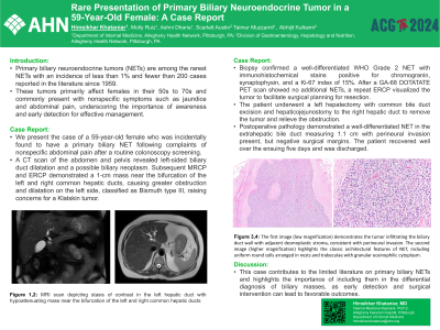
Case Description/Methods: We present the case of a 59-year-old female who was incidentally found to have a primary biliary NET following complaints of nonspecific abdominal pain after a routine colonoscopy screening. A CT scan of the abdomen and pelvis revealed left-sided biliary duct dilatation and a possible biliary neoplasm. Subsequent MRCP and ERCP demonstrated a 1-cm mass near the bifurcation of the left and right common hepatic ducts, causing greater obstruction and dilatation on the left side, classified as Bismuth type III, raising concerns for a Klatskin tumor. Biopsy confirmed a well-differentiated WHO Grade 2 NET with immunohistochemical stains positive for chromogranin, synaptophysin, and a Ki-67 index of 15%. After a GA-68 DOTATATE PET scan showed no additional NETs, a repeat ERCP visualized the tumor to facilitate surgical planning for resection. The patient underwent a left hepatectomy with common bile duct excision and hepaticojejunostomy to the right hepatic duct to remove the tumor and relieve the obstruction. Postoperative pathology demonstrated a well-differentiated NET in the extrahepatic bile duct measuring 1.1 cm with perineural invasion present, but negative surgical margins. The patient recovered well over the ensuing five days and was discharged.
Discussion: This case contributes to the limited literature on primary biliary NETs and highlights the importance of including them in the differential diagnosis of biliary masses, as early detection and surgical intervention can lead to favorable outcomes.

Disclosures:
Himsikhar Khataniar, MD1, Molly Ruiz, BS, MS2, Ashni Dharia, MD1, Scarlett Austin, DO1, Taimur S. Muzammil, MD1, Abhijit Kulkarni, MD3. P0106 - Rare Presentation of Primary Biliary Neuroendocrine Tumor in a 59-Year-Old Female: A Case Report, ACG 2024 Annual Scientific Meeting Abstracts. Philadelphia, PA: American College of Gastroenterology.
1Allegheny General Hospital, Pittsburgh, PA; 2Drexel University College of Medicine, Pittsburgh, PA; 3Allegheny Center for Digestive Health, Pittsburgh, PA
Introduction: Primary biliary neuroendocrine tumors (NETs) are among the rarest NETs with an incidence of less than 1% and fewer than 200 cases reported in the literature since 1959. These tumors primarily affect females in their 50s to 70s and commonly present with nonspecific symptoms such as jaundice and abdominal pain, underscoring the importance of awareness and early detection for effective management.
Case Description/Methods: We present the case of a 59-year-old female who was incidentally found to have a primary biliary NET following complaints of nonspecific abdominal pain after a routine colonoscopy screening. A CT scan of the abdomen and pelvis revealed left-sided biliary duct dilatation and a possible biliary neoplasm. Subsequent MRCP and ERCP demonstrated a 1-cm mass near the bifurcation of the left and right common hepatic ducts, causing greater obstruction and dilatation on the left side, classified as Bismuth type III, raising concerns for a Klatskin tumor. Biopsy confirmed a well-differentiated WHO Grade 2 NET with immunohistochemical stains positive for chromogranin, synaptophysin, and a Ki-67 index of 15%. After a GA-68 DOTATATE PET scan showed no additional NETs, a repeat ERCP visualized the tumor to facilitate surgical planning for resection. The patient underwent a left hepatectomy with common bile duct excision and hepaticojejunostomy to the right hepatic duct to remove the tumor and relieve the obstruction. Postoperative pathology demonstrated a well-differentiated NET in the extrahepatic bile duct measuring 1.1 cm with perineural invasion present, but negative surgical margins. The patient recovered well over the ensuing five days and was discharged.
Discussion: This case contributes to the limited literature on primary biliary NETs and highlights the importance of including them in the differential diagnosis of biliary masses, as early detection and surgical intervention can lead to favorable outcomes.

Figure: Figure 1, 2: MRI scan depicting stasis of contrast in the left hepatic duct with hypoattenuating mass near the bifurcation of the left and right common hepatic ducts
Disclosures:
Himsikhar Khataniar indicated no relevant financial relationships.
Molly Ruiz indicated no relevant financial relationships.
Ashni Dharia indicated no relevant financial relationships.
Scarlett Austin indicated no relevant financial relationships.
Taimur Muzammil indicated no relevant financial relationships.
Abhijit Kulkarni indicated no relevant financial relationships.
Himsikhar Khataniar, MD1, Molly Ruiz, BS, MS2, Ashni Dharia, MD1, Scarlett Austin, DO1, Taimur S. Muzammil, MD1, Abhijit Kulkarni, MD3. P0106 - Rare Presentation of Primary Biliary Neuroendocrine Tumor in a 59-Year-Old Female: A Case Report, ACG 2024 Annual Scientific Meeting Abstracts. Philadelphia, PA: American College of Gastroenterology.
