Sunday Poster Session
Category: Biliary/Pancreas
P0114 - Unmasking a Rare Culprit of Ascending Cholangitis
Sunday, October 27, 2024
3:30 PM - 7:00 PM ET
Location: Exhibit Hall E

Has Audio

Yasmin O. Ali, MBBS
Hennepin Healthcare
Minneapolis, MN
Presenting Author(s)
Yasmin O. Ali, MBBS1, Spencer Goble, MD2, Ahmad Malli, MD1, Yuri Hanada, MD1
1Hennepin Healthcare, Minneapolis, MN; 2University of Minnesota, Minneapolis, MN
Introduction: Biliary ascariasis is rare in the United States (US) and can lead to serious consequences including biliary obstruction, cholangitis, cholecystitis, and acute pancreatitis. This case report highlights the challenges in the diagnosis and management of biliary ascariasis.
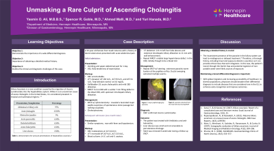
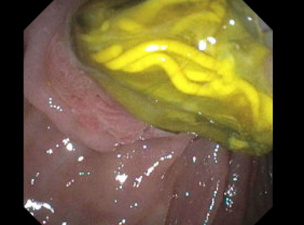
Case Description/Methods: A 34-year-old female from South America with a history of latent tuberculosis presented with acute abdominal pain and vomiting. Vital signs were stable. Physical examination revealed right upper quadrant abdominal tenderness. Liver chemistries revealed alkaline phosphatase (AP) 130 IU/L, alanine transaminase of 93 IU/L, and aspartate aminotransferase (AST) 61 IU/L. Total bilirubin was within normal limits. Gallbladder ultrasound findings were consistent with acute cholecystitis, as well as mild dilatation of the common bile duct (CBD). Subsequent magnetic resonance cholangiopancreatography showed a 9 mm CBD with a subtle 7 mm filling defect in the distal CBD with upstream intrahepatic biliary dilatation. Therefore, she underwent endoscopic retrograde cholangiopancreatography (ERCP), which demonstrated a lacerated major papilla suspicious of spontaneous stone passage, but otherwise nothing was swept from the duct after uncomplicated sphincterotomy was completed. Cholecystectomy was performed and the patient was discharged with complete symptom resolution. However, one month later, she was readmitted with similar symptoms, fever and hypotension. Labs were notable for AP 247 IU/L, ALT 213 IU/L, and leukocytosis 12.5 k/cmm. Due to concern for post-surgical complication, CT scan was performed and showed a 3.2 cm left lobe liver abscess. Repeat ERCP revealed extensive parasitic worm burden at the papillary orifice. Ductal sweeping extracted multiple worms, including a 7 in worm from the main bile duct. A biliary stent was placed. Pathology confirmed Ascaris lumbricoides. The patient was treated with an extended course of antibiotics and an anti-helminthic agent.
Discussion: Ascaris lumbricoides is an uncommon cause of acute cholangitis in the US. However, clinical suspicion should be maintained especially in patients originating from or traveling through endemic regions. In this case, an exposure history revealed transit through the Darién Gap with suspected non-potable water ingestion. With global migration and accessibility of healthcare access for diverse populations, physicians should anticipate the unexpected and broaden their differential diagnosis to include diseases that are considered rare in the US.

Disclosures:
Yasmin O. Ali, MBBS1, Spencer Goble, MD2, Ahmad Malli, MD1, Yuri Hanada, MD1. P0114 - Unmasking a Rare Culprit of Ascending Cholangitis, ACG 2024 Annual Scientific Meeting Abstracts. Philadelphia, PA: American College of Gastroenterology.
1Hennepin Healthcare, Minneapolis, MN; 2University of Minnesota, Minneapolis, MN
Introduction: Biliary ascariasis is rare in the United States (US) and can lead to serious consequences including biliary obstruction, cholangitis, cholecystitis, and acute pancreatitis. This case report highlights the challenges in the diagnosis and management of biliary ascariasis.
Case Description/Methods: A 34-year-old female from South America with a history of latent tuberculosis presented with acute abdominal pain and vomiting. Vital signs were stable. Physical examination revealed right upper quadrant abdominal tenderness. Liver chemistries revealed alkaline phosphatase (AP) 130 IU/L, alanine transaminase of 93 IU/L, and aspartate aminotransferase (AST) 61 IU/L. Total bilirubin was within normal limits. Gallbladder ultrasound findings were consistent with acute cholecystitis, as well as mild dilatation of the common bile duct (CBD). Subsequent magnetic resonance cholangiopancreatography showed a 9 mm CBD with a subtle 7 mm filling defect in the distal CBD with upstream intrahepatic biliary dilatation. Therefore, she underwent endoscopic retrograde cholangiopancreatography (ERCP), which demonstrated a lacerated major papilla suspicious of spontaneous stone passage, but otherwise nothing was swept from the duct after uncomplicated sphincterotomy was completed. Cholecystectomy was performed and the patient was discharged with complete symptom resolution. However, one month later, she was readmitted with similar symptoms, fever and hypotension. Labs were notable for AP 247 IU/L, ALT 213 IU/L, and leukocytosis 12.5 k/cmm. Due to concern for post-surgical complication, CT scan was performed and showed a 3.2 cm left lobe liver abscess. Repeat ERCP revealed extensive parasitic worm burden at the papillary orifice. Ductal sweeping extracted multiple worms, including a 7 in worm from the main bile duct. A biliary stent was placed. Pathology confirmed Ascaris lumbricoides. The patient was treated with an extended course of antibiotics and an anti-helminthic agent.
Discussion: Ascaris lumbricoides is an uncommon cause of acute cholangitis in the US. However, clinical suspicion should be maintained especially in patients originating from or traveling through endemic regions. In this case, an exposure history revealed transit through the Darién Gap with suspected non-potable water ingestion. With global migration and accessibility of healthcare access for diverse populations, physicians should anticipate the unexpected and broaden their differential diagnosis to include diseases that are considered rare in the US.

Figure: Endoscopic retrograde cholangiopancreatography demonstrated the presence of parasites in the major duodenal papilla
Disclosures:
Yasmin Ali indicated no relevant financial relationships.
Spencer Goble indicated no relevant financial relationships.
Ahmad Malli indicated no relevant financial relationships.
Yuri Hanada indicated no relevant financial relationships.
Yasmin O. Ali, MBBS1, Spencer Goble, MD2, Ahmad Malli, MD1, Yuri Hanada, MD1. P0114 - Unmasking a Rare Culprit of Ascending Cholangitis, ACG 2024 Annual Scientific Meeting Abstracts. Philadelphia, PA: American College of Gastroenterology.
