Tuesday Poster Session
Category: Esophagus
P3970 - A Bump in the Esophagus: A Case of an Esophageal Granular Cell Tumor
Tuesday, October 29, 2024
10:30 AM - 4:00 PM ET
Location: Exhibit Hall E

Has Audio

Ahamed Akmal Lebbe
Weill Cornell Medicine-Qatar
Doha, Qatar
Presenting Author(s)
Ahamed Akmal Lebbe, 1, Siddhant Nair, 2, Parvathi Myer, MD3, SriHari Mahadev, MD4, Malik Mushannen, MD5, David Wan, MD4
1Weill Cornell Medicine-Qatar, New York, NY; 2WCM-Q, New York, NY; 3Montefiore Medical Center, New York, NY; 4New York-Presbyterian / Weill Cornell Medical Center, New York, NY; 5New York-Presbyterian / Brooklyn Methodist Hospital, Brooklyn, NY
Introduction: Granular cell tumors are benign, Schwann cell-derived tumors that usually occur on the skin and tongue but can occur anywhere in the body. One rare system to be affected is the gastrointestinal system, and of these, roughly a third occur in the esophagus. The neoplasm is typically asymptomatic but can cause dysphagia in some patients and occasionally reflux symptoms.
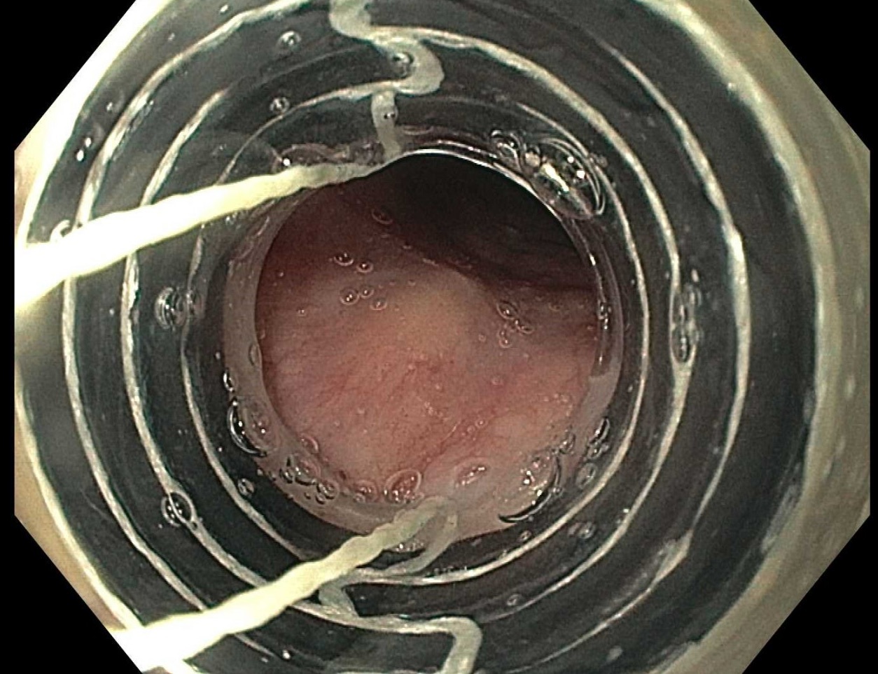
Case Description/Methods: A 48-year-old woman with a history of GERD presented for a colonoscopy and esophagogastroduodenoscopy with a Bravo pH study to evaluate her iron deficiency anemia and reflux symptoms. The colonoscopy found 5 sessile and 3 hyperplastic polyps that were benign. The Bravo pH study showed no pathological levels of acid reflux and EGD showed a regular Z line 40 cm from the incisors without signs of esophagitis. However, a 4 mm submucosal nodule was found in the upper third of the esophagus. An endoscopic ultrasound was performed to further evaluate the nodule. The EUS showed a hypoechoic, 4 mm wide and 3 mm thick lesion with well-defined borders and an intact interface between the nodule and adjacent tissue. The nodule appeared to originate from the submucosa. It was endoscopically resected using cap-assisted band ligation with complete margins ( figure 1 ). Pathology revealed a granular cell tumor.
Discussion: Granular cell tumors of the esophagus are the second most common stromal tumors in the esophagus, therefore being familiar with the endoscopic / EUS appearance and clinical management is important. GCTs appear as a mass with a white / yellow discoloration with normal epithelium. Differential diagnosis includes leiomyomas, stromal cell tumors, lipomas, and schwannomas. Most patients with granular cell tumors of the esophagus present with dysphagia but our patient's was discovered incidentally during her evaluation for acid reflux. Her nodule was found in the upper esophagus which is the rarest location for this kind of tumor with the distal esophagus being more common. Given its size and location, it is doubtful it was related to the acid reflux symptoms. Even when asymptomatic, granular cell tumors have been reported to have a 2-3% chance of malignant transformation, so they should generally be resected. Given their rare occurrence, histologic diagnosis is required. Guidance on surveillance is scant but given the lack of dysplastic findings and complete resection, no surveillance is planned.

Disclosures:
Ahamed Akmal Lebbe, 1, Siddhant Nair, 2, Parvathi Myer, MD3, SriHari Mahadev, MD4, Malik Mushannen, MD5, David Wan, MD4. P3970 - A Bump in the Esophagus: A Case of an Esophageal Granular Cell Tumor, ACG 2024 Annual Scientific Meeting Abstracts. Philadelphia, PA: American College of Gastroenterology.
1Weill Cornell Medicine-Qatar, New York, NY; 2WCM-Q, New York, NY; 3Montefiore Medical Center, New York, NY; 4New York-Presbyterian / Weill Cornell Medical Center, New York, NY; 5New York-Presbyterian / Brooklyn Methodist Hospital, Brooklyn, NY
Introduction: Granular cell tumors are benign, Schwann cell-derived tumors that usually occur on the skin and tongue but can occur anywhere in the body. One rare system to be affected is the gastrointestinal system, and of these, roughly a third occur in the esophagus. The neoplasm is typically asymptomatic but can cause dysphagia in some patients and occasionally reflux symptoms.
Case Description/Methods: A 48-year-old woman with a history of GERD presented for a colonoscopy and esophagogastroduodenoscopy with a Bravo pH study to evaluate her iron deficiency anemia and reflux symptoms. The colonoscopy found 5 sessile and 3 hyperplastic polyps that were benign. The Bravo pH study showed no pathological levels of acid reflux and EGD showed a regular Z line 40 cm from the incisors without signs of esophagitis. However, a 4 mm submucosal nodule was found in the upper third of the esophagus. An endoscopic ultrasound was performed to further evaluate the nodule. The EUS showed a hypoechoic, 4 mm wide and 3 mm thick lesion with well-defined borders and an intact interface between the nodule and adjacent tissue. The nodule appeared to originate from the submucosa. It was endoscopically resected using cap-assisted band ligation with complete margins ( figure 1 ). Pathology revealed a granular cell tumor.
Discussion: Granular cell tumors of the esophagus are the second most common stromal tumors in the esophagus, therefore being familiar with the endoscopic / EUS appearance and clinical management is important. GCTs appear as a mass with a white / yellow discoloration with normal epithelium. Differential diagnosis includes leiomyomas, stromal cell tumors, lipomas, and schwannomas. Most patients with granular cell tumors of the esophagus present with dysphagia but our patient's was discovered incidentally during her evaluation for acid reflux. Her nodule was found in the upper esophagus which is the rarest location for this kind of tumor with the distal esophagus being more common. Given its size and location, it is doubtful it was related to the acid reflux symptoms. Even when asymptomatic, granular cell tumors have been reported to have a 2-3% chance of malignant transformation, so they should generally be resected. Given their rare occurrence, histologic diagnosis is required. Guidance on surveillance is scant but given the lack of dysplastic findings and complete resection, no surveillance is planned.

Figure: Figure 1: Approach to lesion with cap
Disclosures:
Ahamed Akmal Lebbe indicated no relevant financial relationships.
Siddhant Nair indicated no relevant financial relationships.
Parvathi Myer indicated no relevant financial relationships.
SriHari Mahadev: Boston Scientific – Consultant. Conmed – Consultant.
Malik Mushannen indicated no relevant financial relationships.
David Wan indicated no relevant financial relationships.
Ahamed Akmal Lebbe, 1, Siddhant Nair, 2, Parvathi Myer, MD3, SriHari Mahadev, MD4, Malik Mushannen, MD5, David Wan, MD4. P3970 - A Bump in the Esophagus: A Case of an Esophageal Granular Cell Tumor, ACG 2024 Annual Scientific Meeting Abstracts. Philadelphia, PA: American College of Gastroenterology.
