Tuesday Poster Session
Category: Esophagus
P4010 - A Case of Esophageal-Bronchial Fistula Presenting as Dysphagia
Tuesday, October 29, 2024
10:30 AM - 4:00 PM ET
Location: Exhibit Hall E

Has Audio

Nickolas Underhill, DO
Baylor Scott & White Medical Center
Temple, TX
Presenting Author(s)
Nickolas Underhill, DO, Alexis Bejcek, MD, Tengfei Wang, MD, Lisa Lopez, MD, Niket Sonpal, MD
Baylor Scott & White Medical Center, Temple, TX
Introduction: Dysphagia is defined by difficulty swallowing and can have several causes which can include malignancy. Complications of malignancy include the creation of fistulas. A fistula is an abnormal connection between two parts of the body. Patients may present with symptoms including dysphagia, weight loss, hemoptysis, or fever. Our rare case highlights a patient with dysphagia who had a previously normal esophagogastroduodenoscopy (EGD) who was found to have a new esophageal mass with an unexpected finding of an esophageal-bronchial fistula.
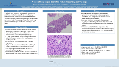
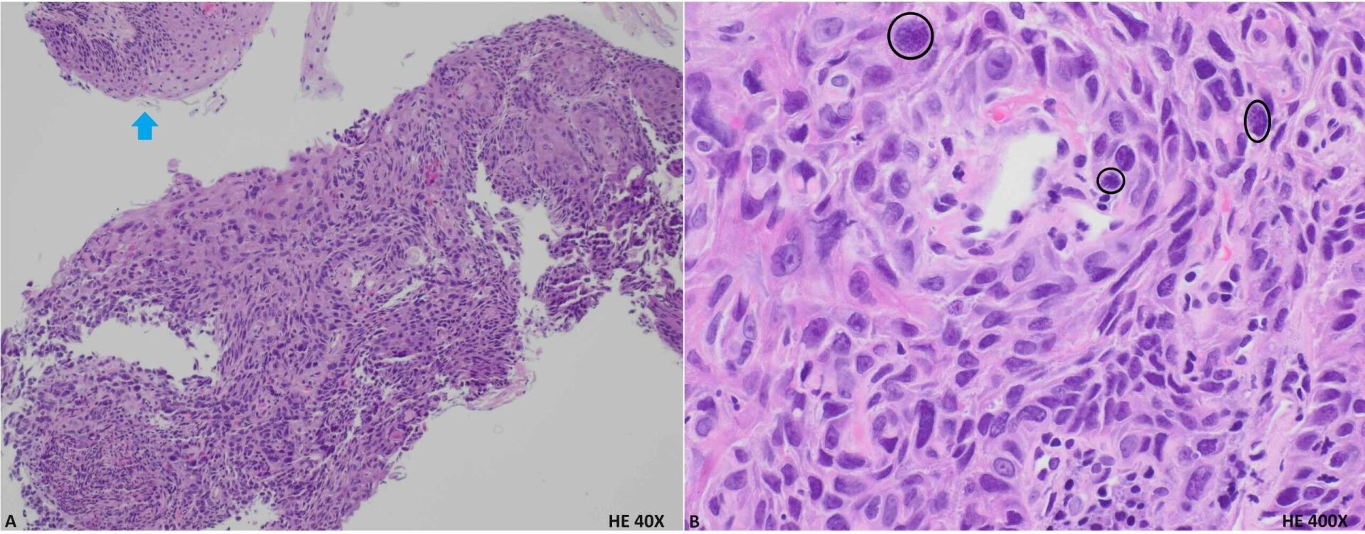
Case Description/Methods: A 60-year-old male presented with dysphagia. The dysphagia started with solids and later included liquids. Additional symptoms included fever, productive cough, hoarseness, and weight loss. EGD several months earlier revealed a normal-appearing esophagus. Imaging showed evidence of aspiration pneumonia and antibiotics were started. Speech and language pathology identified pharyngeal dysphagia. ENT team completed flexible laryngoscopy showing left vocal cord paralysis. CT scan of his neck and chest identified left vocal cord paralysis and a mid-esophageal mass with fistulous connection of the esophagus and the trachea/bronchus among other findings. He underwent bronchoscopy with endobronchial ultrasound-guided transbronchial needle aspiration of station 4R and 7 lymph nodes, balloon dilation of left mainstem followed by Y stent placement by pulmonology and EGD with biopsy by GI. Findings included a 95-100% obstruction of the left mainstem. After balloon dilation and stent placement, there was 0-5% residual obstruction. Rapid on-site evaluation was concerning for squamous cell carcinoma. EGD revealed an esophageal mass causing significant obstruction. Biopsies were obtained. Gastrojejunostomy tube was placed and he was started on tube feeds. He was clinically stable to be discharged home with close follow-up. Final pathology results from the esophageal mass revealed superficial tangential fragments of mostly reactive squamous epithelium with patchy at least high-grade dysplasia.
Discussion: Esophageal-bronchial fistulas may be related to an underlying mass. Causes of these fistulas may include foreign bodies, complications of endoscopic procedures, congenital formation, esophageal diverticula, and malignancy. Our case highlights progressive dysphagia in the setting of vocal cord paralysis with concern for nerve involvement and a new esophageal mass. It also encompasses the importance of interdisciplinary action between several specialties.

Disclosures:
Nickolas Underhill, DO, Alexis Bejcek, MD, Tengfei Wang, MD, Lisa Lopez, MD, Niket Sonpal, MD. P4010 - A Case of Esophageal-Bronchial Fistula Presenting as Dysphagia, ACG 2024 Annual Scientific Meeting Abstracts. Philadelphia, PA: American College of Gastroenterology.
Baylor Scott & White Medical Center, Temple, TX
Introduction: Dysphagia is defined by difficulty swallowing and can have several causes which can include malignancy. Complications of malignancy include the creation of fistulas. A fistula is an abnormal connection between two parts of the body. Patients may present with symptoms including dysphagia, weight loss, hemoptysis, or fever. Our rare case highlights a patient with dysphagia who had a previously normal esophagogastroduodenoscopy (EGD) who was found to have a new esophageal mass with an unexpected finding of an esophageal-bronchial fistula.
Case Description/Methods: A 60-year-old male presented with dysphagia. The dysphagia started with solids and later included liquids. Additional symptoms included fever, productive cough, hoarseness, and weight loss. EGD several months earlier revealed a normal-appearing esophagus. Imaging showed evidence of aspiration pneumonia and antibiotics were started. Speech and language pathology identified pharyngeal dysphagia. ENT team completed flexible laryngoscopy showing left vocal cord paralysis. CT scan of his neck and chest identified left vocal cord paralysis and a mid-esophageal mass with fistulous connection of the esophagus and the trachea/bronchus among other findings. He underwent bronchoscopy with endobronchial ultrasound-guided transbronchial needle aspiration of station 4R and 7 lymph nodes, balloon dilation of left mainstem followed by Y stent placement by pulmonology and EGD with biopsy by GI. Findings included a 95-100% obstruction of the left mainstem. After balloon dilation and stent placement, there was 0-5% residual obstruction. Rapid on-site evaluation was concerning for squamous cell carcinoma. EGD revealed an esophageal mass causing significant obstruction. Biopsies were obtained. Gastrojejunostomy tube was placed and he was started on tube feeds. He was clinically stable to be discharged home with close follow-up. Final pathology results from the esophageal mass revealed superficial tangential fragments of mostly reactive squamous epithelium with patchy at least high-grade dysplasia.
Discussion: Esophageal-bronchial fistulas may be related to an underlying mass. Causes of these fistulas may include foreign bodies, complications of endoscopic procedures, congenital formation, esophageal diverticula, and malignancy. Our case highlights progressive dysphagia in the setting of vocal cord paralysis with concern for nerve involvement and a new esophageal mass. It also encompasses the importance of interdisciplinary action between several specialties.

Figure: Figure 1: Esophagus biopsy shows at least high-grade dysplasia. A: Blue arrow points to relatively normal esophageal squamous mucosa. The tissue below has at least high-grade dysplasia. B: Higher magnification shows the cellular atypia and mitotic figures (black circles).
Disclosures:
Nickolas Underhill indicated no relevant financial relationships.
Alexis Bejcek indicated no relevant financial relationships.
Tengfei Wang indicated no relevant financial relationships.
Lisa Lopez indicated no relevant financial relationships.
Niket Sonpal indicated no relevant financial relationships.
Nickolas Underhill, DO, Alexis Bejcek, MD, Tengfei Wang, MD, Lisa Lopez, MD, Niket Sonpal, MD. P4010 - A Case of Esophageal-Bronchial Fistula Presenting as Dysphagia, ACG 2024 Annual Scientific Meeting Abstracts. Philadelphia, PA: American College of Gastroenterology.
