Tuesday Poster Session
Category: Esophagus
P4012 - Acute Phlegmonous Esophagitis: A Rare Case Report
Tuesday, October 29, 2024
10:30 AM - 4:00 PM ET
Location: Exhibit Hall E

Has Audio

Sanjana S. Nelogal, MBBS
JJM Medical College
Davanagere, Karnataka, India
Presenting Author(s)
Award: Presidential Poster Award
Bala Sai Teja Nuthalapati, MBBS1, Srinivasulu Dudyala, MBBS, MD, DM2, Fnu Rukhayya, MBBS3, Lamees Kaukab, MBBS4, Manognya G, MBBS5, Sanjana S. Nelogal, MBBS6, Aakansha Maria. Rajeev, MBBS7
1Maheshwara Medical College, Hyderabad, Telangana, India; 2Sri Sri Holistic Hospitals, Hyderabad, Telangana, India; 3Dr. VRK Women's Medical College, Hyderabad, Telangana, India; 4Deccan College of Medical Sciences, Hyderabad, Telangana, India; 5Mamata Academy of Medical Sciences, Hyderabad, Telangana, India; 6JJM Medical College, Davanagere, Karnataka, India; 7SDM College of Medical Sciences and Hospital, Dharwad, Karnataka, India
Introduction: Acute phlegmonous esophagitis (APE) is a rare but fatal infection of the submucosa and muscularis of the esophagus. The exact pathogenesis is unclear but risk factors include immunosuppression, alcoholism, uncontrolled diabetes mellitus, old age, and malnutrition. Most patients present with chest pain and dysphagia but rapidly deteriorate to septic shock and death if not treated early and aggressively. Herein, we report a case of APE in a 35-year-old previously healthy man.
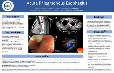
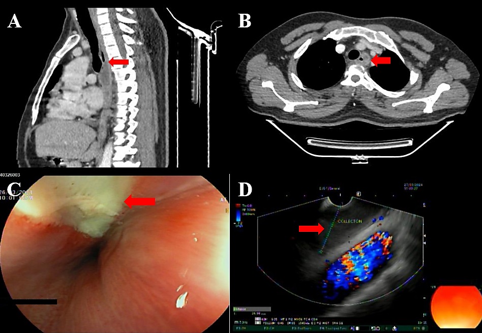
Case Description/Methods: A 35-year-old male presented to the emergency department with worsening chest and throat pain, dysphagia, and odynophagia for a day. He had 1 episode of vomiting but no other symptoms. He has no other medical conditions and doesn't use tobacco, alcohol or other illicit drugs. The physical examination was unremarkable except for mild fever. Esophagogastroduodenoscopy showed a large extrinsic impression on the esophageal wall extending 28-35cm from the incisors. An ulcer with purulent discharge was visible on the esophageal wall at 25cm from the incisors. The patient had antral gastritis with normal duodenum. Esophageal USG demonstrated collection in the submucosal layer of the esophagus. Ulcer discharge culture yielded Pseudomonas aeruginosa. Contrast-enhanced CT of the chest revealed an eccentric long segment, rim enhancing collection along the left lateral and posterior walls of the esophagus throughout the whole length with maximum thickness at mid esophagus sparing gastroesophageal junction suggestive of APE. The patient was treated with intravenous sulbactam, cefoperazone, Meropenem, and Metronidazole for 4 days followed by endoscopic incision and drainage of pus. The patient responded well and had no pus discharge on day 5 endoscopy. The patient was discharged on day 6 with oral cephalosporin and levofloxacin.
Discussion: APE is a rapidly progressive fatal condition. The common causative pathogens include Staphylococcus, Streptococcus, Klebsiella, E.coli, H. influenzae, and Proteus. Pseudomonas causing APE is extremely rare in an immunocompetent person. Except for mild asymptomatic antral gastritis, our patient did not have any known risk factors posing a challenge in the diagnosis. Early diagnosis and treatment are critical to avoid complications like esophageal perforation and mediastinitis. Severe cases may require surgical intervention. This case highlights the importance of considering APE as a differential diagnosis in a patient presenting with chest pain and dysphagia.

Disclosures:
Bala Sai Teja Nuthalapati, MBBS1, Srinivasulu Dudyala, MBBS, MD, DM2, Fnu Rukhayya, MBBS3, Lamees Kaukab, MBBS4, Manognya G, MBBS5, Sanjana S. Nelogal, MBBS6, Aakansha Maria. Rajeev, MBBS7. P4012 - Acute Phlegmonous Esophagitis: A Rare Case Report, ACG 2024 Annual Scientific Meeting Abstracts. Philadelphia, PA: American College of Gastroenterology.
Bala Sai Teja Nuthalapati, MBBS1, Srinivasulu Dudyala, MBBS, MD, DM2, Fnu Rukhayya, MBBS3, Lamees Kaukab, MBBS4, Manognya G, MBBS5, Sanjana S. Nelogal, MBBS6, Aakansha Maria. Rajeev, MBBS7
1Maheshwara Medical College, Hyderabad, Telangana, India; 2Sri Sri Holistic Hospitals, Hyderabad, Telangana, India; 3Dr. VRK Women's Medical College, Hyderabad, Telangana, India; 4Deccan College of Medical Sciences, Hyderabad, Telangana, India; 5Mamata Academy of Medical Sciences, Hyderabad, Telangana, India; 6JJM Medical College, Davanagere, Karnataka, India; 7SDM College of Medical Sciences and Hospital, Dharwad, Karnataka, India
Introduction: Acute phlegmonous esophagitis (APE) is a rare but fatal infection of the submucosa and muscularis of the esophagus. The exact pathogenesis is unclear but risk factors include immunosuppression, alcoholism, uncontrolled diabetes mellitus, old age, and malnutrition. Most patients present with chest pain and dysphagia but rapidly deteriorate to septic shock and death if not treated early and aggressively. Herein, we report a case of APE in a 35-year-old previously healthy man.
Case Description/Methods: A 35-year-old male presented to the emergency department with worsening chest and throat pain, dysphagia, and odynophagia for a day. He had 1 episode of vomiting but no other symptoms. He has no other medical conditions and doesn't use tobacco, alcohol or other illicit drugs. The physical examination was unremarkable except for mild fever. Esophagogastroduodenoscopy showed a large extrinsic impression on the esophageal wall extending 28-35cm from the incisors. An ulcer with purulent discharge was visible on the esophageal wall at 25cm from the incisors. The patient had antral gastritis with normal duodenum. Esophageal USG demonstrated collection in the submucosal layer of the esophagus. Ulcer discharge culture yielded Pseudomonas aeruginosa. Contrast-enhanced CT of the chest revealed an eccentric long segment, rim enhancing collection along the left lateral and posterior walls of the esophagus throughout the whole length with maximum thickness at mid esophagus sparing gastroesophageal junction suggestive of APE. The patient was treated with intravenous sulbactam, cefoperazone, Meropenem, and Metronidazole for 4 days followed by endoscopic incision and drainage of pus. The patient responded well and had no pus discharge on day 5 endoscopy. The patient was discharged on day 6 with oral cephalosporin and levofloxacin.
Discussion: APE is a rapidly progressive fatal condition. The common causative pathogens include Staphylococcus, Streptococcus, Klebsiella, E.coli, H. influenzae, and Proteus. Pseudomonas causing APE is extremely rare in an immunocompetent person. Except for mild asymptomatic antral gastritis, our patient did not have any known risk factors posing a challenge in the diagnosis. Early diagnosis and treatment are critical to avoid complications like esophageal perforation and mediastinitis. Severe cases may require surgical intervention. This case highlights the importance of considering APE as a differential diagnosis in a patient presenting with chest pain and dysphagia.

Figure: Figure A depicts the sagittal view of chest CT with eccentric long segment rim enhancing collection along the left lateral and posterior walls of the esophagus throughout the whole length with maximum thickness at mid esophagus. Figure B depicts axial view of chest CT with hypodense collection in the esophageal wall. Figure C depicts endoscopy of the esophagus showing ulcer with pus discharge. Figure D depicts endoscopic ultrasound revealing collection in the esophageal wall.
Disclosures:
Bala Sai Teja Nuthalapati indicated no relevant financial relationships.
Srinivasulu Dudyala indicated no relevant financial relationships.
Fnu Rukhayya indicated no relevant financial relationships.
Lamees Kaukab indicated no relevant financial relationships.
Manognya G indicated no relevant financial relationships.
Sanjana Nelogal indicated no relevant financial relationships.
Aakansha Rajeev indicated no relevant financial relationships.
Bala Sai Teja Nuthalapati, MBBS1, Srinivasulu Dudyala, MBBS, MD, DM2, Fnu Rukhayya, MBBS3, Lamees Kaukab, MBBS4, Manognya G, MBBS5, Sanjana S. Nelogal, MBBS6, Aakansha Maria. Rajeev, MBBS7. P4012 - Acute Phlegmonous Esophagitis: A Rare Case Report, ACG 2024 Annual Scientific Meeting Abstracts. Philadelphia, PA: American College of Gastroenterology.

