Tuesday Poster Session
Category: Esophagus
P4035 - A Case of the Bright Blue Esophagus: A Rare Endoscopic and Histopathologic Finding
Tuesday, October 29, 2024
10:30 AM - 4:00 PM ET
Location: Exhibit Hall E

Has Audio

Martin Ankamah, MBA
University of St. Thomas Houston
Houston, TX
Presenting Author(s)
Martin Ankamah, MBA1, Robert S.. Aaron, MD2, Stacey Longo, MD3
1University of St. Thomas Houston, Houston, TX; 2Allied Digestive Health (Middlesex Monmouth Gastroenterology), Freehold, NJ; 3Allied Digestive Health, West Long Branch, NJ
Introduction: Esophageal parakeratosis (EP) is an uncommon endoscopic finding usually characterized by linear plaques, mucosal thickenings with rings and furrows, or discrete flat elevated lesions. These findings can appear similar to candida esophagitis, eosinophilic esophagitis, and neoplastic lesions like squamous cell cancer or dysplasia. To date, there are no reported cases of EP progressing to dysplasia or cancer. The morphology of EP shows incomplete keratinization of the esophageal epithelium and compact squamous epithelium with retained pyknotic nuclei. Other studies have reported that EP could be associated with ethanol exposure, vitamin B12 deficiency, zinc deficiency, alcohol cirrhosis, and tylosis. The etiology, clinical significance, and management are not well defined. Thus, the objective of this case report is to add to the current knowledge on esophageal parakeratosis.
Case Description/Methods: A 76-year-old woman with a past medical history significant for hypertension, dyslipidemia, and migraines was referred for evaluation of intermittent dysphagia to solid foods of two years duration without associated reflux. She had two normal swallow evaluations prior to referral but never had an upper endoscopy.
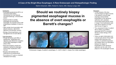
Upper endoscopy demonstrated a bright blue substance coating much of the esophagus which limited visualization of the esophageal mucosa. No obvious stricture was identified. Biopsies demonstrated mild dyskeratosis and parakeratosis, reactive atypia and were reviewed at Cleveland Clinic. Mild antral erythema was noted; the duodenum was normal. Biopsies taken of the stomach demonstrated mild chronic gastritis without evidence of H. pylori or intestinal metaplasia.
Discussion: Further analysis of the blue mucosal plaques (images A and B) in the patient’s mid and distal esophagus demonstrated keratinization of the squamous epithelium (pink cytoplasm surrounding purple pyknotic nuclei in image C) and dyskeratosis (pink layer with an absence of nuclei near the top of image C). This unexpected finding in a patient with no history of conditions associated with EP such as vitamin deficiencies or significant past history of alcohol consumption is consistent with the unclear etiology of this condition. Follow up upper endoscopies over a reasonable period of time might enable further learning on this condition, its potential progression, and whether there is an association between the degree of keratinization of the squamous epithelium and the color of the plaques on the esophageal mucosa.

Disclosures:
Martin Ankamah, MBA1, Robert S.. Aaron, MD2, Stacey Longo, MD3. P4035 - A Case of the Bright Blue Esophagus: A Rare Endoscopic and Histopathologic Finding, ACG 2024 Annual Scientific Meeting Abstracts. Philadelphia, PA: American College of Gastroenterology.
1University of St. Thomas Houston, Houston, TX; 2Allied Digestive Health (Middlesex Monmouth Gastroenterology), Freehold, NJ; 3Allied Digestive Health, West Long Branch, NJ
Introduction: Esophageal parakeratosis (EP) is an uncommon endoscopic finding usually characterized by linear plaques, mucosal thickenings with rings and furrows, or discrete flat elevated lesions. These findings can appear similar to candida esophagitis, eosinophilic esophagitis, and neoplastic lesions like squamous cell cancer or dysplasia. To date, there are no reported cases of EP progressing to dysplasia or cancer. The morphology of EP shows incomplete keratinization of the esophageal epithelium and compact squamous epithelium with retained pyknotic nuclei. Other studies have reported that EP could be associated with ethanol exposure, vitamin B12 deficiency, zinc deficiency, alcohol cirrhosis, and tylosis. The etiology, clinical significance, and management are not well defined. Thus, the objective of this case report is to add to the current knowledge on esophageal parakeratosis.
Case Description/Methods: A 76-year-old woman with a past medical history significant for hypertension, dyslipidemia, and migraines was referred for evaluation of intermittent dysphagia to solid foods of two years duration without associated reflux. She had two normal swallow evaluations prior to referral but never had an upper endoscopy.
Upper endoscopy demonstrated a bright blue substance coating much of the esophagus which limited visualization of the esophageal mucosa. No obvious stricture was identified. Biopsies demonstrated mild dyskeratosis and parakeratosis, reactive atypia and were reviewed at Cleveland Clinic. Mild antral erythema was noted; the duodenum was normal. Biopsies taken of the stomach demonstrated mild chronic gastritis without evidence of H. pylori or intestinal metaplasia.
Discussion: Further analysis of the blue mucosal plaques (images A and B) in the patient’s mid and distal esophagus demonstrated keratinization of the squamous epithelium (pink cytoplasm surrounding purple pyknotic nuclei in image C) and dyskeratosis (pink layer with an absence of nuclei near the top of image C). This unexpected finding in a patient with no history of conditions associated with EP such as vitamin deficiencies or significant past history of alcohol consumption is consistent with the unclear etiology of this condition. Follow up upper endoscopies over a reasonable period of time might enable further learning on this condition, its potential progression, and whether there is an association between the degree of keratinization of the squamous epithelium and the color of the plaques on the esophageal mucosa.

Figure: Endoscopic images of patient’s esophagus: A. mid B. distal. C biopsy from distal esophagus
Disclosures:
Martin Ankamah indicated no relevant financial relationships.
Robert Aaron indicated no relevant financial relationships.
Stacey Longo indicated no relevant financial relationships.
Martin Ankamah, MBA1, Robert S.. Aaron, MD2, Stacey Longo, MD3. P4035 - A Case of the Bright Blue Esophagus: A Rare Endoscopic and Histopathologic Finding, ACG 2024 Annual Scientific Meeting Abstracts. Philadelphia, PA: American College of Gastroenterology.
