Tuesday Poster Session
Category: Esophagus
P4040 - Esophagitis Dessicans Masquerading as a Foreign Object
Tuesday, October 29, 2024
10:30 AM - 4:00 PM ET
Location: Exhibit Hall E

Has Audio
- ZI
Zohaib Ijaz, MD
University of Florida College of Medicine
Gainesville, FL
Presenting Author(s)
Zohaib Ijaz, MD1, Bishal Paudel, MD2, Aleksey Novikov, MD1
1University of Florida College of Medicine, Gainesville, FL; 2University of Florida, Gainesville, FL
Introduction: Esophagitis dissecans superficialis (EDS) is a rare and a self-limiting condition that is characterized by sloughing of the esophageal mucosa. Presenting symptoms include dysphagia, odynophagia or expulsion of the esophageal lining in vomitus or stool. Etiology can include medications (bisphosphonates/NSAIDs), infection, idiopathic, irritant ingestion, esophageal motility disorders or autoimmune diseases. Diagnosis is made via gross endoscopic findings and/or pathology. Endoscopic findings show mucosal layer separation with linear cracks, white plaques and circumferential breaks. Histology shows evidence of epithelium sloughing and parakeratosis.
Case Description/Methods: We report a case of a 68-year-old female with past medical history of abdominal aortic aneurysm with repair, type II diabetes mellitus, end stage kidney disease on dialysis, hypertension, hyperlipidemia, Escherichia coli bacteremia and methicillin-resistant staphylococcus aureus endocarditis who was admitted to the hospital with confusion for hypoglycemia and failure to thrive. Gastroenterology service was consulted for melena. Esophagogastroduodenoscopy (EGD) showed what appeared as a white-green sleeve of non-organic material in the middle third and lower third part of the esophagus. It was not removed and an additional EGD was requested from an interventional GI service. Repeat EGD demonstrated similar findings. EMR technique was used to remove what appeared to be an embedded tough white-green membrane that looked like part of a vinyl glove. Surprisingly, pathology later resulted as squamous epithelium with detached keratin debris and pill fragment material, consistent with esophagitis dissecans superficialis. Further EGDs were not attempted as the patient opted for hospice care due to her other acute on chronic conditions.
Discussion: EDS is a benign condition that can sometimes be difficult to identify. Our case demonstrates a scenario where EDS was initially misdiagnosed as a foreign body. Although it's mostly self-limiting, it is very important to keep EDS in our differential to avoid unnecessary workup and procedures for patients.

Disclosures:
Zohaib Ijaz, MD1, Bishal Paudel, MD2, Aleksey Novikov, MD1. P4040 - Esophagitis Dessicans Masquerading as a Foreign Object, ACG 2024 Annual Scientific Meeting Abstracts. Philadelphia, PA: American College of Gastroenterology.
1University of Florida College of Medicine, Gainesville, FL; 2University of Florida, Gainesville, FL
Introduction: Esophagitis dissecans superficialis (EDS) is a rare and a self-limiting condition that is characterized by sloughing of the esophageal mucosa. Presenting symptoms include dysphagia, odynophagia or expulsion of the esophageal lining in vomitus or stool. Etiology can include medications (bisphosphonates/NSAIDs), infection, idiopathic, irritant ingestion, esophageal motility disorders or autoimmune diseases. Diagnosis is made via gross endoscopic findings and/or pathology. Endoscopic findings show mucosal layer separation with linear cracks, white plaques and circumferential breaks. Histology shows evidence of epithelium sloughing and parakeratosis.
Case Description/Methods: We report a case of a 68-year-old female with past medical history of abdominal aortic aneurysm with repair, type II diabetes mellitus, end stage kidney disease on dialysis, hypertension, hyperlipidemia, Escherichia coli bacteremia and methicillin-resistant staphylococcus aureus endocarditis who was admitted to the hospital with confusion for hypoglycemia and failure to thrive. Gastroenterology service was consulted for melena. Esophagogastroduodenoscopy (EGD) showed what appeared as a white-green sleeve of non-organic material in the middle third and lower third part of the esophagus. It was not removed and an additional EGD was requested from an interventional GI service. Repeat EGD demonstrated similar findings. EMR technique was used to remove what appeared to be an embedded tough white-green membrane that looked like part of a vinyl glove. Surprisingly, pathology later resulted as squamous epithelium with detached keratin debris and pill fragment material, consistent with esophagitis dissecans superficialis. Further EGDs were not attempted as the patient opted for hospice care due to her other acute on chronic conditions.
Discussion: EDS is a benign condition that can sometimes be difficult to identify. Our case demonstrates a scenario where EDS was initially misdiagnosed as a foreign body. Although it's mostly self-limiting, it is very important to keep EDS in our differential to avoid unnecessary workup and procedures for patients.

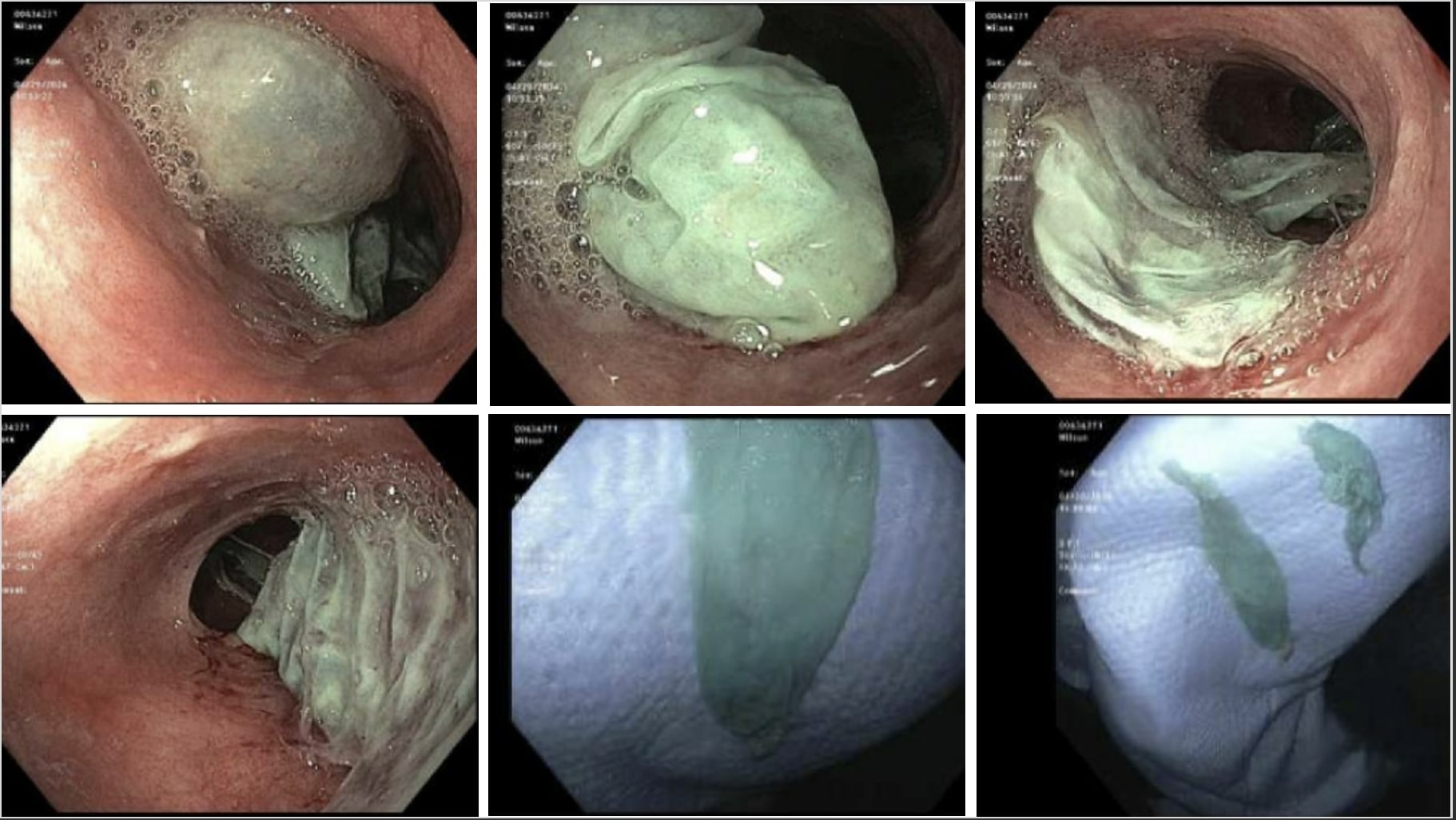
Figure: Top: Esophagitis dissecans in the middle third of the esophagus
Bottom Left: Esophagitis dissecans in the lower third of the esophagus
Bottom Middle/Right: Removed esophagitis dissecans
Bottom Left: Esophagitis dissecans in the lower third of the esophagus
Bottom Middle/Right: Removed esophagitis dissecans
Disclosures:
Zohaib Ijaz indicated no relevant financial relationships.
Bishal Paudel indicated no relevant financial relationships.
Aleksey Novikov indicated no relevant financial relationships.
Zohaib Ijaz, MD1, Bishal Paudel, MD2, Aleksey Novikov, MD1. P4040 - Esophagitis Dessicans Masquerading as a Foreign Object, ACG 2024 Annual Scientific Meeting Abstracts. Philadelphia, PA: American College of Gastroenterology.
