Tuesday Poster Session
Category: General Endoscopy
P4087 - Comparison of Red Dichromatic Imaging With Standard White Light Imaging in Hemostatic Treatment During Endoscopic Interventions: A Systematic Review and Meta-Analysis
Tuesday, October 29, 2024
10:30 AM - 4:00 PM ET
Location: Exhibit Hall E

Has Audio
- SK
Shahryar Khan, MD
University of Kansas
Overland Park, KS
Presenting Author(s)
Shahryar Khan, MD1, Aamer Ahmad, MBBS2, Shehzad Alam Khan, MBBS3, Abdullah Saud, 2, Mashal Alam Khan, MBBS4, Yousaf Zafar, MD5, Usama Ali, MBBS6
1University of Kansas, Overland Park, KS; 2Khyber Medical University, Peshawar, North-West Frontier, Pakistan; 3Peshawar, North-West Frontier, Pakistan; 4Khyber Medical University, Overland Park, KS; 5University of Mississippi Medical Center, Madison, MS; 6Pak International Medical College Peshawar, Mardan, North-West Frontier, Pakistan
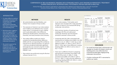
Introduction: It is often difficult to perform endoscopic hemostasis with conventional white light imaging (WLI) for gastrointestinal bleeding when there is blood pooling at the bleeding points. Red dichromatic imaging or dual red imaging (DRI), a novel image-enhanced endoscopy technology, has the potential to improve the visibility of deeper vessels and bleeding points during endoscopic procedures. We aimed to examine the usefulness of DRI in comparison with WLI for endoscopic hemostasis and assessing the visibility of bleeding points.
Methods: We systematically searched Medline, and Embase from inception till Sep 15,2023. The outcomes of interest in our meta-analysis were Visibility score (defined as four numeric values where 1 corresponds to poor visibility and 4 easily detectable), Color difference (defined by CIELAB system), hemostasis time (seconds), and total procedure time (minutes). The random effects model was used to calculate the Standardized Mean difference (SMD) with the corresponding 95% confidence intervals of our desired outcomes. A p value of < 0.05 was considered statistically significant. Heterogeneity was assessed using the I2 Index. Qualities of studies were assessed using the ROBINS-1 tool. Data analysis was performed using the Revman software (version 5.4.1).
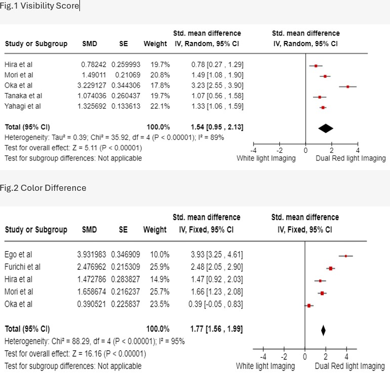
Results: In our meta-analysis, 7 full studies and 4 abstracts were included with a total population of 1645, of which 51.2% received DRI during endoscopic interventions. Most common location of treatment was esophagus (34.5%), and submucosal endoscopic dissection was the frequent procedure assessed in these articles. Comparing with WLI, DRI is associated with improvement of Visibility Score (SMD 1.54, 95% CI 0.95 to 2.13, I2 = 89%, P < 0.05, Fig.1), Color difference (SMD 1.77, 95% CI 1.56 to 1.99, I2 = 95%, P < 0.05, Fig.2), Color difference in oozing bleeding (SMD 2.14, 95% CI 1.63 to 2.66, I2 = 0%, P < 0.05). There was no difference found in comparing both groups for hemostasis time (SMD -0.46, 95% CI 0.93 to 0.01, I2 = 86%, P = 0.06), procedure time (SMD -0.33, 95% CI -0.81 to 0.16, I2 = 78%, P = 0.18), and Color difference in gushing bleeding (SMD 2.97, 95% CI -1.16 to 7.10, I2 = 96%, P = 0.16).
Discussion: Our study found that Dual or dichromatic red imaging can visualize the deeper vessels and bleeding points more effectively than conventional white light imaging during endoscopic interventions. A further multicenter RCT is warranted to confirm our results.

Disclosures:
Shahryar Khan, MD1, Aamer Ahmad, MBBS2, Shehzad Alam Khan, MBBS3, Abdullah Saud, 2, Mashal Alam Khan, MBBS4, Yousaf Zafar, MD5, Usama Ali, MBBS6. P4087 - Comparison of Red Dichromatic Imaging With Standard White Light Imaging in Hemostatic Treatment During Endoscopic Interventions: A Systematic Review and Meta-Analysis, ACG 2024 Annual Scientific Meeting Abstracts. Philadelphia, PA: American College of Gastroenterology.
1University of Kansas, Overland Park, KS; 2Khyber Medical University, Peshawar, North-West Frontier, Pakistan; 3Peshawar, North-West Frontier, Pakistan; 4Khyber Medical University, Overland Park, KS; 5University of Mississippi Medical Center, Madison, MS; 6Pak International Medical College Peshawar, Mardan, North-West Frontier, Pakistan
Introduction: It is often difficult to perform endoscopic hemostasis with conventional white light imaging (WLI) for gastrointestinal bleeding when there is blood pooling at the bleeding points. Red dichromatic imaging or dual red imaging (DRI), a novel image-enhanced endoscopy technology, has the potential to improve the visibility of deeper vessels and bleeding points during endoscopic procedures. We aimed to examine the usefulness of DRI in comparison with WLI for endoscopic hemostasis and assessing the visibility of bleeding points.
Methods: We systematically searched Medline, and Embase from inception till Sep 15,2023. The outcomes of interest in our meta-analysis were Visibility score (defined as four numeric values where 1 corresponds to poor visibility and 4 easily detectable), Color difference (defined by CIELAB system), hemostasis time (seconds), and total procedure time (minutes). The random effects model was used to calculate the Standardized Mean difference (SMD) with the corresponding 95% confidence intervals of our desired outcomes. A p value of < 0.05 was considered statistically significant. Heterogeneity was assessed using the I2 Index. Qualities of studies were assessed using the ROBINS-1 tool. Data analysis was performed using the Revman software (version 5.4.1).
Results: In our meta-analysis, 7 full studies and 4 abstracts were included with a total population of 1645, of which 51.2% received DRI during endoscopic interventions. Most common location of treatment was esophagus (34.5%), and submucosal endoscopic dissection was the frequent procedure assessed in these articles. Comparing with WLI, DRI is associated with improvement of Visibility Score (SMD 1.54, 95% CI 0.95 to 2.13, I2 = 89%, P < 0.05, Fig.1), Color difference (SMD 1.77, 95% CI 1.56 to 1.99, I2 = 95%, P < 0.05, Fig.2), Color difference in oozing bleeding (SMD 2.14, 95% CI 1.63 to 2.66, I2 = 0%, P < 0.05). There was no difference found in comparing both groups for hemostasis time (SMD -0.46, 95% CI 0.93 to 0.01, I2 = 86%, P = 0.06), procedure time (SMD -0.33, 95% CI -0.81 to 0.16, I2 = 78%, P = 0.18), and Color difference in gushing bleeding (SMD 2.97, 95% CI -1.16 to 7.10, I2 = 96%, P = 0.16).
Discussion: Our study found that Dual or dichromatic red imaging can visualize the deeper vessels and bleeding points more effectively than conventional white light imaging during endoscopic interventions. A further multicenter RCT is warranted to confirm our results.

Figure: Forrest plots. Fig.1 Visibility Score and Fig.2 Color Difference
Disclosures:
Shahryar Khan indicated no relevant financial relationships.
Aamer Ahmad indicated no relevant financial relationships.
Shehzad Alam Khan indicated no relevant financial relationships.
Abdullah Saud indicated no relevant financial relationships.
Mashal Alam Khan indicated no relevant financial relationships.
Yousaf Zafar indicated no relevant financial relationships.
Usama Ali indicated no relevant financial relationships.
Shahryar Khan, MD1, Aamer Ahmad, MBBS2, Shehzad Alam Khan, MBBS3, Abdullah Saud, 2, Mashal Alam Khan, MBBS4, Yousaf Zafar, MD5, Usama Ali, MBBS6. P4087 - Comparison of Red Dichromatic Imaging With Standard White Light Imaging in Hemostatic Treatment During Endoscopic Interventions: A Systematic Review and Meta-Analysis, ACG 2024 Annual Scientific Meeting Abstracts. Philadelphia, PA: American College of Gastroenterology.
