Tuesday Poster Session
Category: General Endoscopy
P4123 - Bleeding Duodenal Varices in a Patient with Hepatocellular Cancer - A Challenge in Management
Tuesday, October 29, 2024
10:30 AM - 4:00 PM ET
Location: Exhibit Hall E

Has Audio
.jpg)
Sadia Ansari, MD
University of Oklahoma Health Sciences Center
Oklahoma City, OK
Presenting Author(s)
Sadia Ansari, MD1, Tokunbo Ajayi, MD2
1University of Oklahoma Health Sciences Center, Oklahoma City, OK; 2University of Oklahoma Health Sciences Center, Oklahoma city, OK
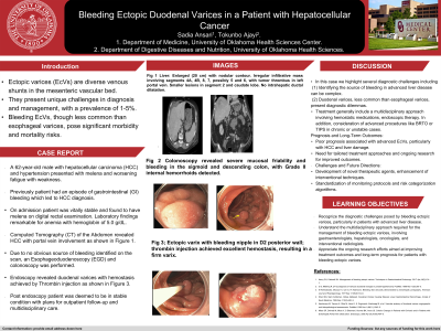
Introduction: Ectopic varices provide special diagnostic and therapeutic issues to providers. This is especially true for patients who have hepatocellular carcinoma (HCC) or other underlying liver diseases. Even though they are uncommon, these varices can cause significant GI bleeding. In cases with HCC, mortality and complications such as embolic stroke is high.
Case Description/Methods: A 62 y/o man with history of hypertension and HCV with sustained virologic response (SVR) after treatment who presented with anemia and recurrent melena after a recent diagnosis of HCC. Physical examination revealed digital rectal examination (DRE) consistent with melena, while vital signs remained stable. Lab showed Hgb of 4.7 g/dL and mild thrombocytosis. Imaging studies, including a computed tomography scan with contrast, demonstrated a large infiltrative liver mass indicative of HCC, with evidence of tumor thrombus invasion into the left portal vein. Subsequent EGD revealed duodenal varices, which were treated with endoscopic gluing and thrombin injection after extensive discussion of risk and benefit during hospitalization and with procedural consent discussion. Following endoscopic procedure, patient's bleeding resolved without needing further transfusion on outpatient follow up.
Discussion: This case of ectopic varices in the duodenum illustrates the range of unconventional locations where varices can arise. These varices are commonly caused by isolated portal hypertension resulting from either portal vein thrombosis or splenic vein thrombosis. The optimal management of bleeding ectopic varices remains unknown; endoscopic techniques in tertiary institutions have been effective. The challenge in this case is that our patient diagnosis of HCC with high Cancer of the Liver Italian Program (CLIP) score of 4 is a significant contraindication to endoscopic procedure. However, without intervention he will bleed to death. Therefore, offering therapy as palliation in an end-of-life situation is appropriate. Interventional radiology procedures such as balloon occluded retrograde transvenous obliteration (BRTO) and transjugular intrahepatic portosystemic shunt (TIPS) are usually contraindicated in this instance due to HCC and tumor thrombus. Complications of endoscopic therapy include brain abscess, digital gangrene, and acute renal failure. This case highlights the need for early detection, patient selection and referral to tertiary institutions that can offer aggressive treatment plans.

Disclosures:
Sadia Ansari, MD1, Tokunbo Ajayi, MD2. P4123 - Bleeding Duodenal Varices in a Patient with Hepatocellular Cancer - A Challenge in Management, ACG 2024 Annual Scientific Meeting Abstracts. Philadelphia, PA: American College of Gastroenterology.
1University of Oklahoma Health Sciences Center, Oklahoma City, OK; 2University of Oklahoma Health Sciences Center, Oklahoma city, OK
Introduction: Ectopic varices provide special diagnostic and therapeutic issues to providers. This is especially true for patients who have hepatocellular carcinoma (HCC) or other underlying liver diseases. Even though they are uncommon, these varices can cause significant GI bleeding. In cases with HCC, mortality and complications such as embolic stroke is high.
Case Description/Methods: A 62 y/o man with history of hypertension and HCV with sustained virologic response (SVR) after treatment who presented with anemia and recurrent melena after a recent diagnosis of HCC. Physical examination revealed digital rectal examination (DRE) consistent with melena, while vital signs remained stable. Lab showed Hgb of 4.7 g/dL and mild thrombocytosis. Imaging studies, including a computed tomography scan with contrast, demonstrated a large infiltrative liver mass indicative of HCC, with evidence of tumor thrombus invasion into the left portal vein. Subsequent EGD revealed duodenal varices, which were treated with endoscopic gluing and thrombin injection after extensive discussion of risk and benefit during hospitalization and with procedural consent discussion. Following endoscopic procedure, patient's bleeding resolved without needing further transfusion on outpatient follow up.
Discussion: This case of ectopic varices in the duodenum illustrates the range of unconventional locations where varices can arise. These varices are commonly caused by isolated portal hypertension resulting from either portal vein thrombosis or splenic vein thrombosis. The optimal management of bleeding ectopic varices remains unknown; endoscopic techniques in tertiary institutions have been effective. The challenge in this case is that our patient diagnosis of HCC with high Cancer of the Liver Italian Program (CLIP) score of 4 is a significant contraindication to endoscopic procedure. However, without intervention he will bleed to death. Therefore, offering therapy as palliation in an end-of-life situation is appropriate. Interventional radiology procedures such as balloon occluded retrograde transvenous obliteration (BRTO) and transjugular intrahepatic portosystemic shunt (TIPS) are usually contraindicated in this instance due to HCC and tumor thrombus. Complications of endoscopic therapy include brain abscess, digital gangrene, and acute renal failure. This case highlights the need for early detection, patient selection and referral to tertiary institutions that can offer aggressive treatment plans.

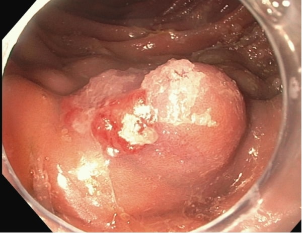
Figure: Duodenal varices post endoscopic treatment
Disclosures:
Sadia Ansari indicated no relevant financial relationships.
Tokunbo Ajayi indicated no relevant financial relationships.
Sadia Ansari, MD1, Tokunbo Ajayi, MD2. P4123 - Bleeding Duodenal Varices in a Patient with Hepatocellular Cancer - A Challenge in Management, ACG 2024 Annual Scientific Meeting Abstracts. Philadelphia, PA: American College of Gastroenterology.
