Tuesday Poster Session
Category: General Endoscopy
P4154 - Down to the Wire: An Esophageal Foreign Body
Tuesday, October 29, 2024
10:30 AM - 4:00 PM ET
Location: Exhibit Hall E

Has Audio
- PS
Pablo Santander, MD
Walter Reed National Military Medical Center
Bethesda, MD
Presenting Author(s)
Pablo Santander, MD, Jared Magee, DO, MPH
Walter Reed National Military Medical Center, Bethesda, MD
Introduction: Foreign body ingestion is a common occurrence. 10-20% of cases need endoscopic intervention. Diagnosis is based on symptoms with imaging. Endoscopy timing is based on the object size and shape. Batteries and sharp objects are of concern due to perforation risk and are to be emergently removed within 2-6 hours. Equipment needed for retrieval depends on the material and shape of the ingested object. We present a case of a wire that was inadvertently ingested and removed with rubber tipped forceps.
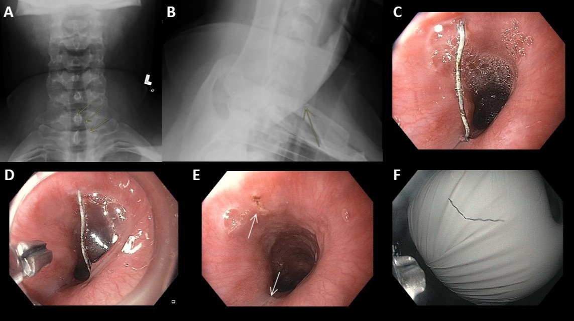
Case Description/Methods: We present the case of a 31-year-old woman with throat pain but able to tolerate secretions. Prior to presenting to the emergency room she was eating tacos. She swallowed and immediately noted severe sharp right-sided throat pain above her clavicle. Vital signs were normal. Her physical exam was notable for right-sided neck tenderness and pain with swallowing. Plain radiographs of the neck identified a thin radiopaque object at the location of pain and was posterior to the trachea on lateral films (Figure 1A and 1B).
The patient consented for endoscopy and was intubated for airway protection in the setting of GLP-1 administration earlier that day. The endoscope was introduced and encountered a wire (Figure 1C) 25 centimeters from the incisors. The wire appeared superficially embedded into the esophageal mucosa on examination and was consistent with her radiographical findings. A cap was placed over the endoscope. The wire was retrieved with rubber tipped forceps (Figure 1D) and pulled into the cap before careful removal (Figure 1E). On re-examination of the esophagus, there was no sign of deep mucosal injury or bleeding (Figure 1F). The patient was extubated and had complete resolution of her pain.
Discussion: Foreign body ingestion is common. Based on patient history, one can surmise the possible object ingested. This knowledge can direct further workup as well as endoscopy timing. Our patient presented with acute sharp throat pain. With ingestion of meat prior to presentation, neck radiography was a reasonable next step (suspected bone ingestion). In our case, a wire-like radiopaque object was identified and directed more emergent endoscopy. Rubber tipped grasping forceps were used to retrieve the wire to minimize slipping. Cap assistance was used to protect the esophagus and upper airway structures. This case highlights the need for detailed history and close examination of imaging, which will dictate endoscopy timing and necessary equipment needed for successful retrieval.

Disclosures:
Pablo Santander, MD, Jared Magee, DO, MPH. P4154 - Down to the Wire: An Esophageal Foreign Body, ACG 2024 Annual Scientific Meeting Abstracts. Philadelphia, PA: American College of Gastroenterology.
Walter Reed National Military Medical Center, Bethesda, MD
Introduction: Foreign body ingestion is a common occurrence. 10-20% of cases need endoscopic intervention. Diagnosis is based on symptoms with imaging. Endoscopy timing is based on the object size and shape. Batteries and sharp objects are of concern due to perforation risk and are to be emergently removed within 2-6 hours. Equipment needed for retrieval depends on the material and shape of the ingested object. We present a case of a wire that was inadvertently ingested and removed with rubber tipped forceps.
Case Description/Methods: We present the case of a 31-year-old woman with throat pain but able to tolerate secretions. Prior to presenting to the emergency room she was eating tacos. She swallowed and immediately noted severe sharp right-sided throat pain above her clavicle. Vital signs were normal. Her physical exam was notable for right-sided neck tenderness and pain with swallowing. Plain radiographs of the neck identified a thin radiopaque object at the location of pain and was posterior to the trachea on lateral films (Figure 1A and 1B).
The patient consented for endoscopy and was intubated for airway protection in the setting of GLP-1 administration earlier that day. The endoscope was introduced and encountered a wire (Figure 1C) 25 centimeters from the incisors. The wire appeared superficially embedded into the esophageal mucosa on examination and was consistent with her radiographical findings. A cap was placed over the endoscope. The wire was retrieved with rubber tipped forceps (Figure 1D) and pulled into the cap before careful removal (Figure 1E). On re-examination of the esophagus, there was no sign of deep mucosal injury or bleeding (Figure 1F). The patient was extubated and had complete resolution of her pain.
Discussion: Foreign body ingestion is common. Based on patient history, one can surmise the possible object ingested. This knowledge can direct further workup as well as endoscopy timing. Our patient presented with acute sharp throat pain. With ingestion of meat prior to presentation, neck radiography was a reasonable next step (suspected bone ingestion). In our case, a wire-like radiopaque object was identified and directed more emergent endoscopy. Rubber tipped grasping forceps were used to retrieve the wire to minimize slipping. Cap assistance was used to protect the esophagus and upper airway structures. This case highlights the need for detailed history and close examination of imaging, which will dictate endoscopy timing and necessary equipment needed for successful retrieval.

Figure: Figure 1A: AP radiograph showing thin radiopaque foreign body.
Figure 1B: Lateral radiograph with foreign body posterior to the trachea.
Figure 1C: Wire encountered in proximal esophagus.
Figure 1D: Cap and rubber tipped forceps used for wire retrieval.
Figure 1E: Mild mucosal injury seen post-retrieval.
Figure 1F: Approximately 3 cm wire
Figure 1B: Lateral radiograph with foreign body posterior to the trachea.
Figure 1C: Wire encountered in proximal esophagus.
Figure 1D: Cap and rubber tipped forceps used for wire retrieval.
Figure 1E: Mild mucosal injury seen post-retrieval.
Figure 1F: Approximately 3 cm wire
Disclosures:
Pablo Santander indicated no relevant financial relationships.
Jared Magee indicated no relevant financial relationships.
Pablo Santander, MD, Jared Magee, DO, MPH. P4154 - Down to the Wire: An Esophageal Foreign Body, ACG 2024 Annual Scientific Meeting Abstracts. Philadelphia, PA: American College of Gastroenterology.
