Tuesday Poster Session
Category: General Endoscopy
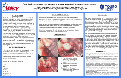
P4155 - Endoscopic Band Ligation as a Temporary Measure to Achieve Hemostasis in Isolated Gastric Varices
Tuesday, October 29, 2024
10:30 AM - 4:00 PM ET
Location: Exhibit Hall E

Has Audio

Chris Pace, DO
Valley Hospital Medical Center
Las Vegas, NV
Presenting Author(s)
Chris Pace, DO, Scott Diamond, DO, Brian Carlson, MD
Valley Hospital Medical Center, Las Vegas, NV
Introduction: Gastric Varices (GV) are dilated submucosal collateral veins caused by portal hypertension. GV have a higher propensity of severe bleed and a higher morbidity depending on size and location. The Sarin classification uses location to describe GV as gastroesophageal varices (GOV) or isolated gastric varices (IGV). The preferred method of endoscopic intervention is sclerotherapy with cyanoacrylate, but its use is limited in the U.S. Band ligation is equally successful in initial hemostasis but is typically used only for GOV due to greater risk of rebleeding. In this case, endoscopic band ligation (EVL) was successfully used as a temporizing measure for a bleeding IGV-1.
Case Description/Methods: A 79 year old male with chronic portal vein and splenic thromboses presents to the hospital with hematemesis and melena for 2 days. Vital signs HR of 88, RR 18, BP 99/51. Labs include Hgb 7.1, Plt 506, BUN 71, creatinine 1.0, Lactic acid 2.7. CT Abdomen/pelvis with contrast showed hepatomegaly, portosystemic varices of the gastric cardia, chronic portal vein occlusion and splenic vein occlusion with venous collaterals. The patient was given 1 unit of pRBCs and initiated on IV pantoprazole 40mg BID and continuous IV octreotide. EGD revealed an actively bleeding isolated GV in the fundus of the stomach (IGV-1). EVL was performed with successful hemostasis. IR, within an hour of EGD, performed a catheter directed venogram of gastric varices with coil embolization. The patient received a total of 3 units pRBCs; hgb stabilized and no other transfusions were required following band therapy and coil embolization.
Discussion: EVL is recommended for GOV-1 lesser curvature GV, but not for non-GOV-1 gastric varices due to rebleeding risk. Studies have shown successful use of cyanoacrylate, but it is not approved by the FDA and can cause complications such as systemic embolism or stroke. Treatment with EVL has varying success rates and is associated with higher risk of rebleeding than cyanoacrylate therapy.
This causes a dilemma with actively bleeding GVs that are not GOV-1, as hemostasis modalities are limited and delay to immediate access for definitive therapy with IR embolization may result in patient harm. EVL of GV may benefit select patients if the rupture site is visualized and the varices can be completely suctioned into the banding cap. This case shows banding to be a viable temporizing measure in select patients to achieve hemostasis which can be life-saving and a bridge to definitive GV obliteration.

Disclosures:
Chris Pace, DO, Scott Diamond, DO, Brian Carlson, MD. P4155 - Endoscopic Band Ligation as a Temporary Measure to Achieve Hemostasis in Isolated Gastric Varices, ACG 2024 Annual Scientific Meeting Abstracts. Philadelphia, PA: American College of Gastroenterology.
Valley Hospital Medical Center, Las Vegas, NV
Introduction: Gastric Varices (GV) are dilated submucosal collateral veins caused by portal hypertension. GV have a higher propensity of severe bleed and a higher morbidity depending on size and location. The Sarin classification uses location to describe GV as gastroesophageal varices (GOV) or isolated gastric varices (IGV). The preferred method of endoscopic intervention is sclerotherapy with cyanoacrylate, but its use is limited in the U.S. Band ligation is equally successful in initial hemostasis but is typically used only for GOV due to greater risk of rebleeding. In this case, endoscopic band ligation (EVL) was successfully used as a temporizing measure for a bleeding IGV-1.
Case Description/Methods: A 79 year old male with chronic portal vein and splenic thromboses presents to the hospital with hematemesis and melena for 2 days. Vital signs HR of 88, RR 18, BP 99/51. Labs include Hgb 7.1, Plt 506, BUN 71, creatinine 1.0, Lactic acid 2.7. CT Abdomen/pelvis with contrast showed hepatomegaly, portosystemic varices of the gastric cardia, chronic portal vein occlusion and splenic vein occlusion with venous collaterals. The patient was given 1 unit of pRBCs and initiated on IV pantoprazole 40mg BID and continuous IV octreotide. EGD revealed an actively bleeding isolated GV in the fundus of the stomach (IGV-1). EVL was performed with successful hemostasis. IR, within an hour of EGD, performed a catheter directed venogram of gastric varices with coil embolization. The patient received a total of 3 units pRBCs; hgb stabilized and no other transfusions were required following band therapy and coil embolization.
Discussion: EVL is recommended for GOV-1 lesser curvature GV, but not for non-GOV-1 gastric varices due to rebleeding risk. Studies have shown successful use of cyanoacrylate, but it is not approved by the FDA and can cause complications such as systemic embolism or stroke. Treatment with EVL has varying success rates and is associated with higher risk of rebleeding than cyanoacrylate therapy.
This causes a dilemma with actively bleeding GVs that are not GOV-1, as hemostasis modalities are limited and delay to immediate access for definitive therapy with IR embolization may result in patient harm. EVL of GV may benefit select patients if the rupture site is visualized and the varices can be completely suctioned into the banding cap. This case shows banding to be a viable temporizing measure in select patients to achieve hemostasis which can be life-saving and a bridge to definitive GV obliteration.

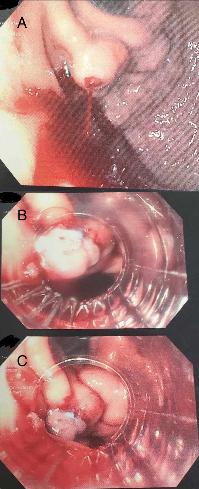
Figure: A. Actively bleeding isolated gastric varix located in the fundus of the stomach (IGV-1).
B. Isolated gastric varix after endoscopic variceal band ligation.
C. Lateral view of isolated gastric varix after endoscopic variceal band ligation.
B. Isolated gastric varix after endoscopic variceal band ligation.
C. Lateral view of isolated gastric varix after endoscopic variceal band ligation.
Disclosures:
Chris Pace indicated no relevant financial relationships.
Scott Diamond indicated no relevant financial relationships.
Brian Carlson indicated no relevant financial relationships.
Chris Pace, DO, Scott Diamond, DO, Brian Carlson, MD. P4155 - Endoscopic Band Ligation as a Temporary Measure to Achieve Hemostasis in Isolated Gastric Varices, ACG 2024 Annual Scientific Meeting Abstracts. Philadelphia, PA: American College of Gastroenterology.
