Tuesday Poster Session
Category: GI Bleeding
P4189 - Overconfident With Over-the-Scope Clips (OTSC)
Tuesday, October 29, 2024
10:30 AM - 4:00 PM ET
Location: Exhibit Hall E

Has Audio
- HS
Hillary C. Smalley, DO
Naval Medical Center San Diego
San Diego, CA
Presenting Author(s)
Hillary C. Smalley, DO, Scott Liu, MD
Naval Medical Center San Diego, San Diego, CA
Introduction: We present a case of an upper gastrointestinal bleed treated with (OTSC) with second look endoscopy findings of hemostasis, intact clip with more than expected healthy appearing mucosa. Subsequent bleeding demonstrated potential flaws and nuance with OTSCs.
Case Description/Methods: A 45-year-old male with history of chronic gastroesophageal reflux disease admitted with hematochezia requiring resuscitation and transfusions. A CT angiogram (CTA) was negative. EGD showed clean-based ulcers in the duodenal bulb and a 6mm ulcer at distal anterior bulb with an oozing visible vessel. Attempts with through the scope clips (TTSC) were unsuccessful due to the inability to close confidently to secure hemostasis due to the ulcer associated fibrotic tissue. An OTSC was deployed with hemostasis. He was placed on PPI IV every 12 hours. He experienced bleeding the following day with repeat CTA notable for an intraluminal 1.5 x 1.4 x 1 cm submucosal soft tissue mass at the area of the OTSC. A second look EGD revealed no blood or heme and intact OTSC with clip created polypoid lesion with visible vessel, but evident hemostasis. A colonoscopy was only remarkable for heme in terminal ileum. On the third EGD, the OTSC created polypoid lesion visible vessel was evidently bleeding. Initial TTCS was placed above the clip and below the visible vessel. TTCS placed over the visible vessel unfortunately malfunctioned and failed to separate requiring removal with tissue that caused the other TTS to dislodge as well. The site was injected with 10 mL of 1:10,000 epinephrine to assist with tamponade/hemostasis. Bipolar coagulation unsuccessful likely secondary to underlying OTSC. Ultimately another OTSC was placed above and near the prior OTSC as the likely site of a feeding vessel with good hemostasis and endoscopic hemostatic powder added for additional coverage.
Discussion: OTSCs are relatively new tools in hemostasis. This case highlights the importance of identification of OTSC unexpected and nuanced findings. More than expected tissue captured by OTSC and normal appearing mucosa on second EGD are indication that active blood flow was still occurring at that area. We recommend on second look endoscopy, if an OTCS created polypoid lesion mucosa is healthy appearing, then there should be a high degree of suspicion for further bleeding and especially if there is a visible vessel. Attempts should be made to further close the vessel or control areas of the feeding vessel.

Disclosures:
Hillary C. Smalley, DO, Scott Liu, MD. P4189 - Overconfident With Over-the-Scope Clips (OTSC), ACG 2024 Annual Scientific Meeting Abstracts. Philadelphia, PA: American College of Gastroenterology.
Naval Medical Center San Diego, San Diego, CA
Introduction: We present a case of an upper gastrointestinal bleed treated with (OTSC) with second look endoscopy findings of hemostasis, intact clip with more than expected healthy appearing mucosa. Subsequent bleeding demonstrated potential flaws and nuance with OTSCs.
Case Description/Methods: A 45-year-old male with history of chronic gastroesophageal reflux disease admitted with hematochezia requiring resuscitation and transfusions. A CT angiogram (CTA) was negative. EGD showed clean-based ulcers in the duodenal bulb and a 6mm ulcer at distal anterior bulb with an oozing visible vessel. Attempts with through the scope clips (TTSC) were unsuccessful due to the inability to close confidently to secure hemostasis due to the ulcer associated fibrotic tissue. An OTSC was deployed with hemostasis. He was placed on PPI IV every 12 hours. He experienced bleeding the following day with repeat CTA notable for an intraluminal 1.5 x 1.4 x 1 cm submucosal soft tissue mass at the area of the OTSC. A second look EGD revealed no blood or heme and intact OTSC with clip created polypoid lesion with visible vessel, but evident hemostasis. A colonoscopy was only remarkable for heme in terminal ileum. On the third EGD, the OTSC created polypoid lesion visible vessel was evidently bleeding. Initial TTCS was placed above the clip and below the visible vessel. TTCS placed over the visible vessel unfortunately malfunctioned and failed to separate requiring removal with tissue that caused the other TTS to dislodge as well. The site was injected with 10 mL of 1:10,000 epinephrine to assist with tamponade/hemostasis. Bipolar coagulation unsuccessful likely secondary to underlying OTSC. Ultimately another OTSC was placed above and near the prior OTSC as the likely site of a feeding vessel with good hemostasis and endoscopic hemostatic powder added for additional coverage.
Discussion: OTSCs are relatively new tools in hemostasis. This case highlights the importance of identification of OTSC unexpected and nuanced findings. More than expected tissue captured by OTSC and normal appearing mucosa on second EGD are indication that active blood flow was still occurring at that area. We recommend on second look endoscopy, if an OTCS created polypoid lesion mucosa is healthy appearing, then there should be a high degree of suspicion for further bleeding and especially if there is a visible vessel. Attempts should be made to further close the vessel or control areas of the feeding vessel.

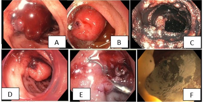
Figure: Image Labels:
A: Duodenal ulcer with visible vessel
B: Over the Scope Clip Padlock
C: Terminal Ileum
D: Prior Padlock Site
E: Second Padlock placed
F: Hemospray application
A: Duodenal ulcer with visible vessel
B: Over the Scope Clip Padlock
C: Terminal Ileum
D: Prior Padlock Site
E: Second Padlock placed
F: Hemospray application
Disclosures:
Hillary Smalley indicated no relevant financial relationships.
Scott Liu indicated no relevant financial relationships.
Hillary C. Smalley, DO, Scott Liu, MD. P4189 - Overconfident With Over-the-Scope Clips (OTSC), ACG 2024 Annual Scientific Meeting Abstracts. Philadelphia, PA: American College of Gastroenterology.
