Tuesday Poster Session
Category: GI Bleeding
P4196 - Malignancy-Induced Hemophilia A in an Elderly Woman
Tuesday, October 29, 2024
10:30 AM - 4:00 PM ET
Location: Exhibit Hall E

Has Audio
- JT
Jefferson Tran
TMC
Sherman, TX
Presenting Author(s)
Marcus Byrd, DO1, David Deuth, DO2, Matthew Ford, 1, Jefferson Tran, 2, Aaron Cernero, DO1, Thomas Tran, MD1
1TMC, Denison, TX; 2TMC, Sherman, TX
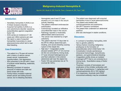
Introduction: Hereditary hemophilia A is an X-linked recessive disorder diagnosed mostly in male children. Acquired hemophilia A (AHA) is a rare autoimmune disorder caused by autoantibodies against coagulation factor VIII. AHA carries an incidence of 1.48 cases per million persons per year. We report a case of acquired hemophilia A secondary to cecal adenocarcinoma which led to near fatal GI bleeding in an elderly woman.
Case Description/Methods: Patient is a 79-year-old woman with no family history of bleeding disorders who presented with 3 days of profuse rectal bleeding, weakness, and fatigue. Bleeding consisted of massive amounts of melena and dark red blood mixed with clots. Hemoglobin was 9. CT scan showed a 4.5 cm mass in the cecum actively bleeding. Patient underwent endovascular embolization to control the bleeding. Colonoscopy revealed an infiltrative circumferential mass in the cecum. Pathology reported invasive adenocarcinoma. The cancer was resected by a right colectomy, and patient was discharged home. She returned 10 days later for diffuse abdominal pain, bloating, and severe bleeding to the point of rupture of the staple line. Hgb was 7. Coagulation profile showed isolated PTT of 123 (normal 22-30) not corrected by mixing. CT scan showed heterogeneous air and fluid collection in the abdominal wall near the staple line. Patient continued to bleed profusely at the surgical site. A bleeding disorder was suspected. Factor VIII was extremely low at 1% (normal 56-140%). Patient was diagnosed with acquired hemophilia A due to cecal adenocarcinoma. She was treated with cyclophosphamide and prednisone, resulting in resolution of bleeding. PTT was normalized. Patient underwent an abdominal washout and was discharged in stable conditions.
Discussion: In contrast to hereditary hemophilia, AHA has no genetic patterns. Malignancies predispose to the formation of autoantibodies which inhibit factor VIII. Patients typically develop extensive bleeding which can be life-threatening. Coagulation profile shows an isolated prolonged PTT not corrected by a mixing study. The quantitative assay of factor VIII is low. Treatment consists of hemostasis management and eradication of the inhibitor with high dose of prednisone (1 mg/kg/day) and cyclophosphamide (50 to 100 mg/day). If no responses, rituximab (anti-CD20 monoclonal antibody) may be considered. Clinicians should become familiar with the prompt recognition, diagnosis, and treatment of AHA as it carries significant risks for morbidity and mortality.
Disclosures:
Marcus Byrd, DO1, David Deuth, DO2, Matthew Ford, 1, Jefferson Tran, 2, Aaron Cernero, DO1, Thomas Tran, MD1. P4196 - Malignancy-Induced Hemophilia A in an Elderly Woman, ACG 2024 Annual Scientific Meeting Abstracts. Philadelphia, PA: American College of Gastroenterology.
1TMC, Denison, TX; 2TMC, Sherman, TX
Introduction: Hereditary hemophilia A is an X-linked recessive disorder diagnosed mostly in male children. Acquired hemophilia A (AHA) is a rare autoimmune disorder caused by autoantibodies against coagulation factor VIII. AHA carries an incidence of 1.48 cases per million persons per year. We report a case of acquired hemophilia A secondary to cecal adenocarcinoma which led to near fatal GI bleeding in an elderly woman.
Case Description/Methods: Patient is a 79-year-old woman with no family history of bleeding disorders who presented with 3 days of profuse rectal bleeding, weakness, and fatigue. Bleeding consisted of massive amounts of melena and dark red blood mixed with clots. Hemoglobin was 9. CT scan showed a 4.5 cm mass in the cecum actively bleeding. Patient underwent endovascular embolization to control the bleeding. Colonoscopy revealed an infiltrative circumferential mass in the cecum. Pathology reported invasive adenocarcinoma. The cancer was resected by a right colectomy, and patient was discharged home. She returned 10 days later for diffuse abdominal pain, bloating, and severe bleeding to the point of rupture of the staple line. Hgb was 7. Coagulation profile showed isolated PTT of 123 (normal 22-30) not corrected by mixing. CT scan showed heterogeneous air and fluid collection in the abdominal wall near the staple line. Patient continued to bleed profusely at the surgical site. A bleeding disorder was suspected. Factor VIII was extremely low at 1% (normal 56-140%). Patient was diagnosed with acquired hemophilia A due to cecal adenocarcinoma. She was treated with cyclophosphamide and prednisone, resulting in resolution of bleeding. PTT was normalized. Patient underwent an abdominal washout and was discharged in stable conditions.
Discussion: In contrast to hereditary hemophilia, AHA has no genetic patterns. Malignancies predispose to the formation of autoantibodies which inhibit factor VIII. Patients typically develop extensive bleeding which can be life-threatening. Coagulation profile shows an isolated prolonged PTT not corrected by a mixing study. The quantitative assay of factor VIII is low. Treatment consists of hemostasis management and eradication of the inhibitor with high dose of prednisone (1 mg/kg/day) and cyclophosphamide (50 to 100 mg/day). If no responses, rituximab (anti-CD20 monoclonal antibody) may be considered. Clinicians should become familiar with the prompt recognition, diagnosis, and treatment of AHA as it carries significant risks for morbidity and mortality.
Disclosures:
Marcus Byrd indicated no relevant financial relationships.
David Deuth indicated no relevant financial relationships.
Matthew Ford indicated no relevant financial relationships.
Jefferson Tran indicated no relevant financial relationships.
Aaron Cernero indicated no relevant financial relationships.
Thomas Tran indicated no relevant financial relationships.
Marcus Byrd, DO1, David Deuth, DO2, Matthew Ford, 1, Jefferson Tran, 2, Aaron Cernero, DO1, Thomas Tran, MD1. P4196 - Malignancy-Induced Hemophilia A in an Elderly Woman, ACG 2024 Annual Scientific Meeting Abstracts. Philadelphia, PA: American College of Gastroenterology.
