Tuesday Poster Session
Category: General Endoscopy
P4145 - A Collaborative Endoscopic-Surgical Approach to Sigmoid Volvulus Caused by Peritoneal Metastases
Tuesday, October 29, 2024
10:30 AM - 4:00 PM ET
Location: Exhibit Hall E

Has Audio

Aabid Mohiuddin, DO
Detroit Medical Center/Wayne State University
Detroit, MI
Presenting Author(s)
Aabid Mohiuddin, DO1, Uzair Khan, BS2, Eric Denha, MD1, Fadi Antaki, MD3, Christine Schad, MD3, Samara Rifkin, MD1
1Detroit Medical Center/Wayne State University, Detroit, MI; 2Wayne State University School of Medicine, Detroit, MI; 3John D. Dingell VA Medical Center, Detroit, MI
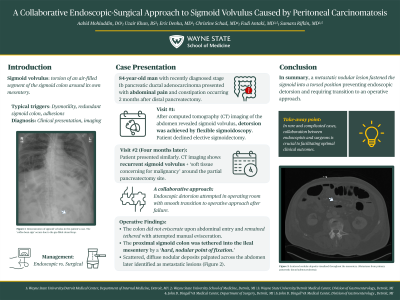
Introduction: Sigmoid volvulus— torsion of an air-filled segment of the sigmoid colon around its own mesentery— is a rare obstructive pathology requiring prompt evaluation. Both endoscopic and surgical interventions are viable depending on the presence of alarm symptoms (i.e., peritonitis). We detail below a rare case of sigmoid volvulus caused by metastatic peritoneal lesions.
Case Description/Methods: An 84-year-old man with recently diagnosed stage 1b pancreatic ductal adenocarcinoma presented with five days of abdominal pain and constipation two months after distal pancreatectomy at an outside institution. Mild distention was noted on abdominal exam; laboratory evaluation was unremarkable. Computed tomography imaging with contrast revealed sigmoid volvulus; after surgical and gastroenterology evaluation, complete detorsion was achieved by flexible sigmoidoscopy with marked symptomatic improvement. The patient declined elective sigmoidectomy.
Four months later, he returned with identical symptoms and similar physical exam and laboratory findings. Imaging revealed recurrent sigmoid volvulus as well as ‘soft tissue concerning for malignancy’ around the partial pancreatectomy site. Symptomatic improvement was achieved with endoscopic decompression, however abdominal x-ray obtained following the procedure showed decompression but ongoing volvulus.
Despite an additional attempt to detorse the sigmoid, the volvulus remained fixed. The endoscopic procedure was performed in the operating room with the colorectal surgeon present, allowing for immediate conversion to an operative approach. Notably, the colon did not eviscerate upon abdominal entry and remained tethered with attempted manual evisceration. The proximal sigmoid colon traversed the midline with its mesentery tethered to the ileal mesentery by a ‘hard, nodular point of fixation.’ Scattered, diffuse nodular deposits later identified as metastatic lesions were palpated across the abdomen. Successful sigmoid colectomy with end colostomy was subsequently performed.
Discussion: Typically, sigmoid volvulus occurs due to redundant sigmoid colon or dysmotility and can be untwisted endoscopically prior to surgery to improve surgical outcomes. In this case, a metastatic nodular lesion acted as a button, fastening the sigmoid into a torsed position preventing endoscopic detorsion. This case highlights the importance of collaboration between endoscopists and surgeons as the seamless transition from endoscopic to operative management facilitated a successful outcome.

Disclosures:
Aabid Mohiuddin, DO1, Uzair Khan, BS2, Eric Denha, MD1, Fadi Antaki, MD3, Christine Schad, MD3, Samara Rifkin, MD1. P4145 - A Collaborative Endoscopic-Surgical Approach to Sigmoid Volvulus Caused by Peritoneal Metastases, ACG 2024 Annual Scientific Meeting Abstracts. Philadelphia, PA: American College of Gastroenterology.
1Detroit Medical Center/Wayne State University, Detroit, MI; 2Wayne State University School of Medicine, Detroit, MI; 3John D. Dingell VA Medical Center, Detroit, MI
Introduction: Sigmoid volvulus— torsion of an air-filled segment of the sigmoid colon around its own mesentery— is a rare obstructive pathology requiring prompt evaluation. Both endoscopic and surgical interventions are viable depending on the presence of alarm symptoms (i.e., peritonitis). We detail below a rare case of sigmoid volvulus caused by metastatic peritoneal lesions.
Case Description/Methods: An 84-year-old man with recently diagnosed stage 1b pancreatic ductal adenocarcinoma presented with five days of abdominal pain and constipation two months after distal pancreatectomy at an outside institution. Mild distention was noted on abdominal exam; laboratory evaluation was unremarkable. Computed tomography imaging with contrast revealed sigmoid volvulus; after surgical and gastroenterology evaluation, complete detorsion was achieved by flexible sigmoidoscopy with marked symptomatic improvement. The patient declined elective sigmoidectomy.
Four months later, he returned with identical symptoms and similar physical exam and laboratory findings. Imaging revealed recurrent sigmoid volvulus as well as ‘soft tissue concerning for malignancy’ around the partial pancreatectomy site. Symptomatic improvement was achieved with endoscopic decompression, however abdominal x-ray obtained following the procedure showed decompression but ongoing volvulus.
Despite an additional attempt to detorse the sigmoid, the volvulus remained fixed. The endoscopic procedure was performed in the operating room with the colorectal surgeon present, allowing for immediate conversion to an operative approach. Notably, the colon did not eviscerate upon abdominal entry and remained tethered with attempted manual evisceration. The proximal sigmoid colon traversed the midline with its mesentery tethered to the ileal mesentery by a ‘hard, nodular point of fixation.’ Scattered, diffuse nodular deposits later identified as metastatic lesions were palpated across the abdomen. Successful sigmoid colectomy with end colostomy was subsequently performed.
Discussion: Typically, sigmoid volvulus occurs due to redundant sigmoid colon or dysmotility and can be untwisted endoscopically prior to surgery to improve surgical outcomes. In this case, a metastatic nodular lesion acted as a button, fastening the sigmoid into a torsed position preventing endoscopic detorsion. This case highlights the importance of collaboration between endoscopists and surgeons as the seamless transition from endoscopic to operative management facilitated a successful outcome.

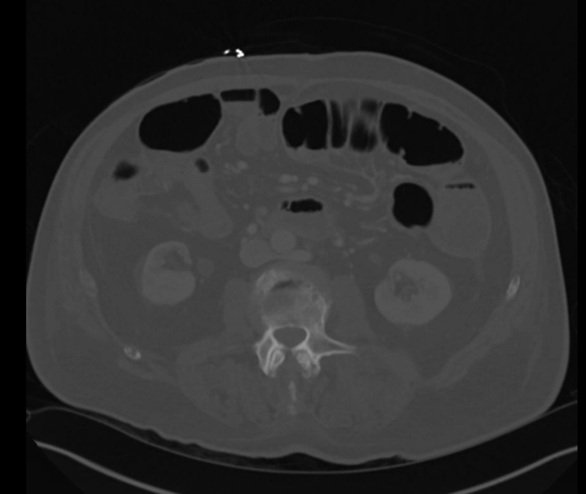
Figure: Computed Tomography (CT) imaging obtained after the operative sigmoidectomy shows the omental metastatic nodules.
Disclosures:
Aabid Mohiuddin indicated no relevant financial relationships.
Uzair Khan indicated no relevant financial relationships.
Eric Denha indicated no relevant financial relationships.
Fadi Antaki indicated no relevant financial relationships.
Christine Schad indicated no relevant financial relationships.
Samara Rifkin indicated no relevant financial relationships.
Aabid Mohiuddin, DO1, Uzair Khan, BS2, Eric Denha, MD1, Fadi Antaki, MD3, Christine Schad, MD3, Samara Rifkin, MD1. P4145 - A Collaborative Endoscopic-Surgical Approach to Sigmoid Volvulus Caused by Peritoneal Metastases, ACG 2024 Annual Scientific Meeting Abstracts. Philadelphia, PA: American College of Gastroenterology.
