Tuesday Poster Session
Category: Esophagus
P3975 - An Incidental Finding: Distal Esophageal Thickening and Middle Mediastinal Venous Malformation With Communicating Vein to the Left Atrium
Tuesday, October 29, 2024
10:30 AM - 4:00 PM ET
Location: Exhibit Hall E

Has Audio

Meghan Cloutier, MD
University at Buffalo
Buffalo, NY
Presenting Author(s)
Meghan Cloutier, MD, Mitchell Edquist, MD, Anthony Khoury, DO, Erin K. Ly, MD, Ramon Rivera, MD, Omar Chohan, DO, Thamer Qaqish, MD
University at Buffalo, Buffalo, NY
Introduction: Distal esophageal thickening is a concerning finding, warranting further workup in the appropriate setting. While distal esophageal thickening is often benign, malignancy does need to be excluded. Unexpected pathology may be discovered, including venous malformations (VMs). Mediastinal VMs are rare with few instances reported. The American College of Radiology Appropriateness Criteria recommends further imaging such as MRI/PET CT for incidental mediastinal masses. Management of these lesions has less clarity.
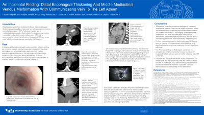
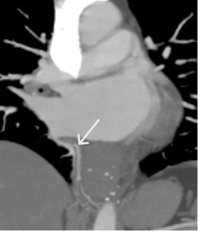
Case Description/Methods: A 50-year-old female underwent coronary calcium scoring. An incidental mass-like thickening of the distal esophagus was seen, further evaluation was recommended. EGD was performed and demonstrated no inflammation or masses, only mucosal discoloration. A CT scan was obtained with near circumferential thickening of the distal one third of the esophagus. Vascular structures were present in the mediastinum which did not enhance during the arterial phase but did in the more delayed phase suggesting venous origin. One tiny vessel along the right lateral margin of the mediastinum demonstrated enhancement in the arterial phase and drained into the left atrium after extending from the retrocrural region. On EUS, the discoloration was once again seen but also revealed the presence of tubal anechoic structures in the lower third of the esophagus with flow on doppler.
Multidisciplinary tumor board decided on no further interventions due to the asymptomatic nature and risks outweighing benefits.
Discussion: EGD is indicated for distal esophageal thickening seen on imaging in the appropriate setting. The American College of Radiology has recommendations based on consensus on the management of thoracic incidental findings; generally these require further characterization with MRI or PET/CT. Mediastinal VMs can be suspected based on the presence of phleboliths and enhancement patterns on contrast-enhanced CT. Additional confirmation can be obtained with endoscopic ultrasound. Malformations of this nature are rare, especially those with a draining vein to the left atrium as in our case which has only been described in the literature twice before.
As imaging volume increases, it is important to follow guidelines regarding follow up with the goal of minimizing patient risk, while maximizing diagnostic yield. There are often not guidelines for these findings and management may vary by case.

Disclosures:
Meghan Cloutier, MD, Mitchell Edquist, MD, Anthony Khoury, DO, Erin K. Ly, MD, Ramon Rivera, MD, Omar Chohan, DO, Thamer Qaqish, MD. P3975 - An Incidental Finding: Distal Esophageal Thickening and Middle Mediastinal Venous Malformation With Communicating Vein to the Left Atrium, ACG 2024 Annual Scientific Meeting Abstracts. Philadelphia, PA: American College of Gastroenterology.
University at Buffalo, Buffalo, NY
Introduction: Distal esophageal thickening is a concerning finding, warranting further workup in the appropriate setting. While distal esophageal thickening is often benign, malignancy does need to be excluded. Unexpected pathology may be discovered, including venous malformations (VMs). Mediastinal VMs are rare with few instances reported. The American College of Radiology Appropriateness Criteria recommends further imaging such as MRI/PET CT for incidental mediastinal masses. Management of these lesions has less clarity.
Case Description/Methods: A 50-year-old female underwent coronary calcium scoring. An incidental mass-like thickening of the distal esophagus was seen, further evaluation was recommended. EGD was performed and demonstrated no inflammation or masses, only mucosal discoloration. A CT scan was obtained with near circumferential thickening of the distal one third of the esophagus. Vascular structures were present in the mediastinum which did not enhance during the arterial phase but did in the more delayed phase suggesting venous origin. One tiny vessel along the right lateral margin of the mediastinum demonstrated enhancement in the arterial phase and drained into the left atrium after extending from the retrocrural region. On EUS, the discoloration was once again seen but also revealed the presence of tubal anechoic structures in the lower third of the esophagus with flow on doppler.
Multidisciplinary tumor board decided on no further interventions due to the asymptomatic nature and risks outweighing benefits.
Discussion: EGD is indicated for distal esophageal thickening seen on imaging in the appropriate setting. The American College of Radiology has recommendations based on consensus on the management of thoracic incidental findings; generally these require further characterization with MRI or PET/CT. Mediastinal VMs can be suspected based on the presence of phleboliths and enhancement patterns on contrast-enhanced CT. Additional confirmation can be obtained with endoscopic ultrasound. Malformations of this nature are rare, especially those with a draining vein to the left atrium as in our case which has only been described in the literature twice before.
As imaging volume increases, it is important to follow guidelines regarding follow up with the goal of minimizing patient risk, while maximizing diagnostic yield. There are often not guidelines for these findings and management may vary by case.

Figure: Coronal contrast-enhanced CT image with MIP (maximum intensity projection) reformatting demonstrates a prominent draining vein (white arrow) communicating to the left atrium.
Disclosures:
Meghan Cloutier indicated no relevant financial relationships.
Mitchell Edquist indicated no relevant financial relationships.
Anthony Khoury indicated no relevant financial relationships.
Erin Ly indicated no relevant financial relationships.
Ramon Rivera indicated no relevant financial relationships.
Omar Chohan indicated no relevant financial relationships.
Thamer Qaqish indicated no relevant financial relationships.
Meghan Cloutier, MD, Mitchell Edquist, MD, Anthony Khoury, DO, Erin K. Ly, MD, Ramon Rivera, MD, Omar Chohan, DO, Thamer Qaqish, MD. P3975 - An Incidental Finding: Distal Esophageal Thickening and Middle Mediastinal Venous Malformation With Communicating Vein to the Left Atrium, ACG 2024 Annual Scientific Meeting Abstracts. Philadelphia, PA: American College of Gastroenterology.

