Tuesday Poster Session
Category: Esophagus
P3977 - Esophageal Fordyce Spots (EFS) - Ectopic Esophageal Sebaceous Glands (EESG)
Tuesday, October 29, 2024
10:30 AM - 4:00 PM ET
Location: Exhibit Hall E

Has Audio
- SB
Shreya Bollu
St. Louis University
Colonia, NJ
Presenting Author(s)
Shreya Bollu, 1, Sugandha Bollu, 1, Elizabeth Eapen, MD2, Metin Taskin, MD2, Janardhan Bollu, MD, FACG3, Sita Chokhavatia, MD, MACG4
1St. Louis University, Colonia, NJ; 2The Valley Hospital, Ridgewood, NJ; 3St. Joseph's University Medical Center, Edison, NJ; 4The Valley Hospital, Paramus, NJ
Introduction: Sebaceous glands are located adjacent to hair follicles throughout the mid-dermis, except on palmar surfaces of hand and soles of feet. Sebaceous glands can be located on ectopic sites including the vermilion border and oral mucosa of the upper lip, buccal mucosa, mandibular retro molar pad, tonsillar fossa and genitals. Esophageal Fordyce Spots (EFS) are benign visible sebaceous glands seen on the lips, gums, inner cheeks and the genitals.
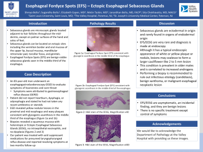
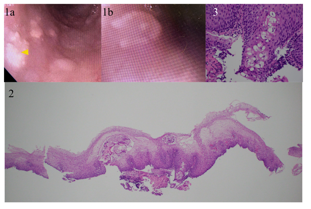
Case Description/Methods: A 83-year-old man underwent an esophagogastroduodenoscopy (EGD) for evaluation of his symptoms of hoarseness and sore throat that were attributed to gastroesophageal reflux disease (GERD). He did not report heartburn or dysphagia or odynophagia and stated he had not taken any recent antibiotics or steroids. EGD revealed yellow nodular lesions in the proximal and mid esophagus and waxy plaques consistent with glycogenic acanthosis in the middle third of the esophagus (Figure 1a and 1b). Biopsies revealed a squamous mucosa with heterotopic or Ectopic Esophageal Sebaceous Glands (EESG), no intraepithelial eosinophils, and no dysplasia (Figures 2 and 3). Our patient was treated with acid suppressant medications for presumed laryngopharyngeal reflux disease and reported resolving symptoms at two months follow up.
Discussion: Sebaceous glands are ectodermal in origin and rarely found in organs of endodermal origin. EFS/EESG are very rare and diagnosis is made at endoscopy. Although it has a typical endoscopic appearance of white or yellow plaque or nodule, lesions may coalesce to form a larger cauliflower like 2 to 5 mm lesion. This condition is prevalent in older males and is correlated to increased androgens. Performing a biopsy is recommended to rule out infectious etiology (candidiasis), benign xanthoma or malignant esophageal neoplastic lesion. EFS/EESG are asymptomatic, an incidental finding, and they are benign lesions and as such, there is no specific treatment.

Disclosures:
Shreya Bollu, 1, Sugandha Bollu, 1, Elizabeth Eapen, MD2, Metin Taskin, MD2, Janardhan Bollu, MD, FACG3, Sita Chokhavatia, MD, MACG4. P3977 - Esophageal Fordyce Spots (EFS) - Ectopic Esophageal Sebaceous Glands (EESG), ACG 2024 Annual Scientific Meeting Abstracts. Philadelphia, PA: American College of Gastroenterology.
1St. Louis University, Colonia, NJ; 2The Valley Hospital, Ridgewood, NJ; 3St. Joseph's University Medical Center, Edison, NJ; 4The Valley Hospital, Paramus, NJ
Introduction: Sebaceous glands are located adjacent to hair follicles throughout the mid-dermis, except on palmar surfaces of hand and soles of feet. Sebaceous glands can be located on ectopic sites including the vermilion border and oral mucosa of the upper lip, buccal mucosa, mandibular retro molar pad, tonsillar fossa and genitals. Esophageal Fordyce Spots (EFS) are benign visible sebaceous glands seen on the lips, gums, inner cheeks and the genitals.
Case Description/Methods: A 83-year-old man underwent an esophagogastroduodenoscopy (EGD) for evaluation of his symptoms of hoarseness and sore throat that were attributed to gastroesophageal reflux disease (GERD). He did not report heartburn or dysphagia or odynophagia and stated he had not taken any recent antibiotics or steroids. EGD revealed yellow nodular lesions in the proximal and mid esophagus and waxy plaques consistent with glycogenic acanthosis in the middle third of the esophagus (Figure 1a and 1b). Biopsies revealed a squamous mucosa with heterotopic or Ectopic Esophageal Sebaceous Glands (EESG), no intraepithelial eosinophils, and no dysplasia (Figures 2 and 3). Our patient was treated with acid suppressant medications for presumed laryngopharyngeal reflux disease and reported resolving symptoms at two months follow up.
Discussion: Sebaceous glands are ectodermal in origin and rarely found in organs of endodermal origin. EFS/EESG are very rare and diagnosis is made at endoscopy. Although it has a typical endoscopic appearance of white or yellow plaque or nodule, lesions may coalesce to form a larger cauliflower like 2 to 5 mm lesion. This condition is prevalent in older males and is correlated to increased androgens. Performing a biopsy is recommended to rule out infectious etiology (candidiasis), benign xanthoma or malignant esophageal neoplastic lesion. EFS/EESG are asymptomatic, an incidental finding, and they are benign lesions and as such, there is no specific treatment.

Figure: Figure 1a: Esophageal Fordyce Spot (EFS)
Figure 1b: Esophageal Fordyce Spot (EFS)
Figure 2: H&E stain of the EESG, Magnification x40
Figure 3: H&E stain of the EESG, Magnification x400
Figure 1b: Esophageal Fordyce Spot (EFS)
Figure 2: H&E stain of the EESG, Magnification x40
Figure 3: H&E stain of the EESG, Magnification x400
Disclosures:
Shreya Bollu indicated no relevant financial relationships.
Sugandha Bollu indicated no relevant financial relationships.
Elizabeth Eapen indicated no relevant financial relationships.
Metin Taskin indicated no relevant financial relationships.
Janardhan Bollu indicated no relevant financial relationships.
Sita Chokhavatia indicated no relevant financial relationships.
Shreya Bollu, 1, Sugandha Bollu, 1, Elizabeth Eapen, MD2, Metin Taskin, MD2, Janardhan Bollu, MD, FACG3, Sita Chokhavatia, MD, MACG4. P3977 - Esophageal Fordyce Spots (EFS) - Ectopic Esophageal Sebaceous Glands (EESG), ACG 2024 Annual Scientific Meeting Abstracts. Philadelphia, PA: American College of Gastroenterology.

