Tuesday Poster Session
Category: Esophagus
P3987 - An Incidental Case of Esophageal Submucosal Gland Duct Adenoma
Tuesday, October 29, 2024
10:30 AM - 4:00 PM ET
Location: Exhibit Hall E

Has Audio
- JM
Jaslyn Maurer, DO
New York-Presbyterian/Queens
Flushing, NY
Presenting Author(s)
Jaslyn Maurer, DO, Hasan Ali, DO, Andrew Schreiner, MD, Syed Hussain, MD
New York-Presbyterian/Queens, Flushing, NY
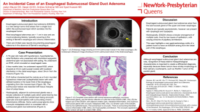
Introduction: Esophageal submucosal gland duct adenoma (ESGDA) is a rare benign tumor that arises from a single duct within the submucosal layer which secretes into the esophageal lumen. Most esophageal adenomas are less than 1 cm in size and are mucin-producing lesions, typically occurring in the setting of Barrett’s esophagus and chronic inflammation. There are limited case reports documenting esophageal adenoma in the absence of Barrett’s esophagus. We report a case of a 76-year-old male with epigastric abdominal pain, who was incidentally found to have an esophageal nodule on esophagogastroduodenoscopy (EGD) later identified as a submucosal gland duct adenoma.
Case Description/Methods: A 76-year-old male with atrial fibrillation presented with intermittent epigastric abdominal pain not associated with eating. He underwent an EGD, which revealed an esophageal mass. A few months later, he underwent repeat EGD, which showed a 8-mm submucosal nodule with localized distribution in the distal esophagus, about 39-cm from the incisors (Figure 1A-B). Endoscopic ultrasound (EUS) further characterized the nodule as a 6-mm rounded hypoechoic intramural (subepithelial) lesion with well-defined borders in the lower third of the esophagus originating from the mucosa layer (Figure 1C-D). The submucosal nodule was resected with tissue margins clear of polyp tissue. Histologically, adjacent to submucosal glands was a lobular collection of multiple cysts which were lined by two epithelial layers, with the luminal layer being oncocytic (granular and eosinophilic) with surrounding chronic inflammatory infiltrate. There was focal papillary architecture. Some submucosal glands showed oncocytic metaplasia which was consistent with a submucosal gland duct adenomas (Figure 1E-H).
Discussion: Esophageal submucosal gland duct adenomas arise from the submucosal gland of the upper and lower esophagus. Patients are typically asymptomatic, however can present with dysphagia and dyspepsia. Histologically, ESGDA shows a lobular arrangement of multiple cysts, lined by two layers of epithelium, with varying amounts of papillary architecture. This case highlights a rare gastroenterology entity of a patient found to have an ESGDA arising from the distal part of the esophagus. Although ESGDA are rare, recognizing these distinct histopathological characteristics is important to create a differential diagnosis, especially because these tumors have similar morphology to their salivary counterparts, such as Warthin tumor of the parotid gland.

Disclosures:
Jaslyn Maurer, DO, Hasan Ali, DO, Andrew Schreiner, MD, Syed Hussain, MD. P3987 - An Incidental Case of Esophageal Submucosal Gland Duct Adenoma, ACG 2024 Annual Scientific Meeting Abstracts. Philadelphia, PA: American College of Gastroenterology.
New York-Presbyterian/Queens, Flushing, NY
Introduction: Esophageal submucosal gland duct adenoma (ESGDA) is a rare benign tumor that arises from a single duct within the submucosal layer which secretes into the esophageal lumen. Most esophageal adenomas are less than 1 cm in size and are mucin-producing lesions, typically occurring in the setting of Barrett’s esophagus and chronic inflammation. There are limited case reports documenting esophageal adenoma in the absence of Barrett’s esophagus. We report a case of a 76-year-old male with epigastric abdominal pain, who was incidentally found to have an esophageal nodule on esophagogastroduodenoscopy (EGD) later identified as a submucosal gland duct adenoma.
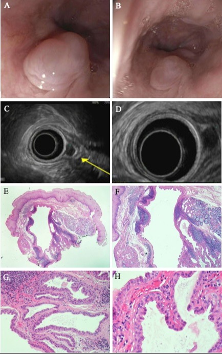
Case Description/Methods: A 76-year-old male with atrial fibrillation presented with intermittent epigastric abdominal pain not associated with eating. He underwent an EGD, which revealed an esophageal mass. A few months later, he underwent repeat EGD, which showed a 8-mm submucosal nodule with localized distribution in the distal esophagus, about 39-cm from the incisors (Figure 1A-B). Endoscopic ultrasound (EUS) further characterized the nodule as a 6-mm rounded hypoechoic intramural (subepithelial) lesion with well-defined borders in the lower third of the esophagus originating from the mucosa layer (Figure 1C-D). The submucosal nodule was resected with tissue margins clear of polyp tissue. Histologically, adjacent to submucosal glands was a lobular collection of multiple cysts which were lined by two epithelial layers, with the luminal layer being oncocytic (granular and eosinophilic) with surrounding chronic inflammatory infiltrate. There was focal papillary architecture. Some submucosal glands showed oncocytic metaplasia which was consistent with a submucosal gland duct adenomas (Figure 1E-H).
Discussion: Esophageal submucosal gland duct adenomas arise from the submucosal gland of the upper and lower esophagus. Patients are typically asymptomatic, however can present with dysphagia and dyspepsia. Histologically, ESGDA shows a lobular arrangement of multiple cysts, lined by two layers of epithelium, with varying amounts of papillary architecture. This case highlights a rare gastroenterology entity of a patient found to have an ESGDA arising from the distal part of the esophagus. Although ESGDA are rare, recognizing these distinct histopathological characteristics is important to create a differential diagnosis, especially because these tumors have similar morphology to their salivary counterparts, such as Warthin tumor of the parotid gland.

Figure: Figure 1: (A,B) Endoscopy image showing an 8-mm submucosal nodule in the distal esophagus. (C,D) Endoscopic Ultrasound image showing an 8-mm submucosal nodule in the distal esophagus. (E) esophageal biopsy showing a complete cross-section of the specimen showing normal squamous epithelium, mucinous submucosal glands, and the lobular collection of cysts that is the ESGDA (H&E, 20x). (F) the largest cyst of the ESGDA, with adjacent smaller cysts, interstitial inflammation, and mucinous submucosal glands with oncocytic metaplasia (H&E, 40x). (G) Smaller cysts of the ESGDA, lined by two epithelial layers. The luminal layer is oncocytic. There is surrounding interstitial chronic inflammation (H&E, 200x). (H) Focal papillary architecture. The oncocytic appearance of the luminal cell layer is evident (H&E, 400x).
Disclosures:
Jaslyn Maurer indicated no relevant financial relationships.
Hasan Ali indicated no relevant financial relationships.
Andrew Schreiner indicated no relevant financial relationships.
Syed Hussain indicated no relevant financial relationships.
Jaslyn Maurer, DO, Hasan Ali, DO, Andrew Schreiner, MD, Syed Hussain, MD. P3987 - An Incidental Case of Esophageal Submucosal Gland Duct Adenoma, ACG 2024 Annual Scientific Meeting Abstracts. Philadelphia, PA: American College of Gastroenterology.
