Tuesday Poster Session
Category: Esophagus
P3988 - Incidental Endoscopic Finding: Esophageal Lymphangioma
Tuesday, October 29, 2024
10:30 AM - 4:00 PM ET
Location: Exhibit Hall E

Has Audio
- JM
Jaslyn Maurer, DO
New York-Presbyterian/Queens
Flushing, NY
Presenting Author(s)
Jaslyn Maurer, DO, Hasan Ali, DO, Jiankun Tong, MD, Syed Hussain, MD
New York-Presbyterian/Queens, Flushing, NY
Introduction: Lymphangiomas are a rare submucosal malformations of the lymphatic system associated with dilated lymphatic vessels, that arise in the neck, axilla, groin, and rarely occur in the gastrointestinal tract, especially in the esophagus. Lymphangiomas are usually asymptomatic and incidentally found on endoscopic evaluation. We present a case of a 56-year old male with atrial fibrillation, gastroesophageal reflux and epigastric abdominal pain, incidentally found to have a 5 mm submucosal nodule seen on esophagogastroduodenoscopy (EGD) and endoscopic ultrasound (EUS) with histological evaluation showing lymphangioma.
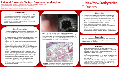
Case Description/Methods: A 57-year old male with paroxysmal atrial fibrillation was being evaluated for gastroesophageal reflux and epigastric abdominal pain. On prior EGD, he was noted to have a distal esophageal lesion. On repeat endoscopic evaluation, there was a 5 mm submucosal nodule in the lower third of the esophagus (about 38 cm from the incisors) (Figure 1A), along with diffuse inflammation of the entire stomach and one non-bleeding 10 mm superficial gastric ulcer on the greater curvature. EUS showed an oval intramural (subepithelial) lesion originating from the deep mucosa (layer 2) measuring up to 3 mm by 2 mm in thickness in the lower third of the esophagus (Figure 1B). No other masses were identified. The endoscope was reinserted and a 17 mm area was resected using band ligator and snare for mucosal resection. Resected tissue margins were examined and clear of polyp tissue and 6 hemostatic clips were successfully placed. Pathology was notable for squamous mucosa showing lymphangioma of the esophagus (Figure 1C). He tolerated the procedure well, and continues to take pantoprazole for gastritis.
Discussion: Lymphangiomas are a benign soft tissue tumor that rarely occurs in the gastrointestinal tract in adults, with about 1% of lymphangiomas originating for the gastrointestinal tract. The most common site within the gastrointestinal tract is the colon, with the esophagus rarely involved. Unlike gastrointestinal mesentery, head or neck lymphangiomas, those that occur in the esophagus tend to occur in adulthood. Most frequently they are diagnosed based on endoscopic evaluation with complete resection, and thus patients have a tendency to have nonspecific symptoms. This case highlights a rare gastroenterology entity of a patient found to have an esophageal lymphangioma arising from the distal portion of the esophagus.

Disclosures:
Jaslyn Maurer, DO, Hasan Ali, DO, Jiankun Tong, MD, Syed Hussain, MD. P3988 - Incidental Endoscopic Finding: Esophageal Lymphangioma, ACG 2024 Annual Scientific Meeting Abstracts. Philadelphia, PA: American College of Gastroenterology.
New York-Presbyterian/Queens, Flushing, NY
Introduction: Lymphangiomas are a rare submucosal malformations of the lymphatic system associated with dilated lymphatic vessels, that arise in the neck, axilla, groin, and rarely occur in the gastrointestinal tract, especially in the esophagus. Lymphangiomas are usually asymptomatic and incidentally found on endoscopic evaluation. We present a case of a 56-year old male with atrial fibrillation, gastroesophageal reflux and epigastric abdominal pain, incidentally found to have a 5 mm submucosal nodule seen on esophagogastroduodenoscopy (EGD) and endoscopic ultrasound (EUS) with histological evaluation showing lymphangioma.
Case Description/Methods: A 57-year old male with paroxysmal atrial fibrillation was being evaluated for gastroesophageal reflux and epigastric abdominal pain. On prior EGD, he was noted to have a distal esophageal lesion. On repeat endoscopic evaluation, there was a 5 mm submucosal nodule in the lower third of the esophagus (about 38 cm from the incisors) (Figure 1A), along with diffuse inflammation of the entire stomach and one non-bleeding 10 mm superficial gastric ulcer on the greater curvature. EUS showed an oval intramural (subepithelial) lesion originating from the deep mucosa (layer 2) measuring up to 3 mm by 2 mm in thickness in the lower third of the esophagus (Figure 1B). No other masses were identified. The endoscope was reinserted and a 17 mm area was resected using band ligator and snare for mucosal resection. Resected tissue margins were examined and clear of polyp tissue and 6 hemostatic clips were successfully placed. Pathology was notable for squamous mucosa showing lymphangioma of the esophagus (Figure 1C). He tolerated the procedure well, and continues to take pantoprazole for gastritis.
Discussion: Lymphangiomas are a benign soft tissue tumor that rarely occurs in the gastrointestinal tract in adults, with about 1% of lymphangiomas originating for the gastrointestinal tract. The most common site within the gastrointestinal tract is the colon, with the esophagus rarely involved. Unlike gastrointestinal mesentery, head or neck lymphangiomas, those that occur in the esophagus tend to occur in adulthood. Most frequently they are diagnosed based on endoscopic evaluation with complete resection, and thus patients have a tendency to have nonspecific symptoms. This case highlights a rare gastroenterology entity of a patient found to have an esophageal lymphangioma arising from the distal portion of the esophagus.

Figure: Figure 1: (A) EGD image showing a 5 mm submucosal nodule with localized distribution found in the lower third of the esophagus. (B) EUS image showing an oval intramural (subepithelial) lesion in the lower third of the esophagus located within the deep mucosa (layer 2) and measured up to 3 mm by 2 mm in thickness. (C) Lymphangioma is located in the submucosa, and it shows dilated spaces lined by flattened endothelium (x 40).
Disclosures:
Jaslyn Maurer indicated no relevant financial relationships.
Hasan Ali indicated no relevant financial relationships.
Jiankun Tong indicated no relevant financial relationships.
Syed Hussain indicated no relevant financial relationships.
Jaslyn Maurer, DO, Hasan Ali, DO, Jiankun Tong, MD, Syed Hussain, MD. P3988 - Incidental Endoscopic Finding: Esophageal Lymphangioma, ACG 2024 Annual Scientific Meeting Abstracts. Philadelphia, PA: American College of Gastroenterology.
