Monday Poster Session
Category: Liver
P3036 - Multifocal Hepatic Small Vessel Neoplasm: A Novelty and Mystery
Monday, October 28, 2024
10:30 AM - 4:00 PM ET
Location: Exhibit Hall E


Sharifeh Almasaid, MD
SUNY Upstate Medical University
Syracuse, NY
Presenting Author(s)
Sharifeh Almasaid, MD1, Dayana Nasr, MD2, Ahmad Nawaz, MD3, Arwa Elsamny, MBBCh3, Savio John, MD, FACG3
1SUNY Upstate Medical University, Johnson City, TN; 2SUNY Upstate Medical University Hospital, Syracuse, NY; 3SUNY Upstate Medical University, Syracuse, NY
Introduction: Hepatic small vessel neoplasm (HSVN) is a rare clinical entity, was first described in 2016. There have only been about thirty cases of HSVN reported so far. It is typically seen in adults with male predominance with no clear symptoms or risk factors. It is generally identified incidentally on imaging as a solitary tumor with variable size, and the diagnosis is confirmed by biopsy and immunohistochemistry staining. HSVN mimics hemangiomas and hepatic angiosarcoma which makes the diagnosis challenging, and yet so far, its potential for malignancy is unknown.
Case Description/Methods: A 66-year-old male who is compound heterozygous for hereditary hemochromatosis (C282Y and H63D mutations), used to work as a carpenter and had occupational exposure to benzene in the past, came to the clinic for evaluation of incidental small indeterminate solid appearing right hepatic hypoechoic nodules. Serological tests for viral hepatitis, iron studies, alpha-fetoprotein, and liver biochemical tests were normal. MRI of the abdomen revealed numerous T2 hyperintense homogenously enhancing right hepatic lesions, the largest measured 1.3* 0.9 cm, and vascular lesions throughout the spleen. A biopsy of the liver lesion was obtained, showing hepatic small vessel neoplasm with features of vascular proliferation. The case was discussed in tumor board. He was not a surgical candidate due to multiple lesions; he was referred to liver transplant center who recommended a surveillance MRI every 3 months to monitor progression.
Discussion: HSVN is a rare vascular tumor and has similar molecular biology to congenital hemangioma and anastomosing hemangioma with GNAQ, GNA11 and GNA14 mutations. It is believed to have a low recurrence rate and minimal potential for malignant transformation. Imaging can show similarities with hepatocellular carcinoma, hepatic angiosarcoma, and atypical vascular tumors, making a biopsy essential for diagnosis. Although HSVN is commonly associated with underlying liver disease, its association between hereditary hemochromatosis and/or exposure to benzene has not been reported yet, and this is the first case of such association. Multifocal disseminated HSVN has been described only once in literature, and to best of our knowledge this is the second reported case involving the liver and the spleen. The current recommendation is to resect the tumor and monitor it with serial imaging in the case of solitary liver lesions.

Disclosures:
Sharifeh Almasaid, MD1, Dayana Nasr, MD2, Ahmad Nawaz, MD3, Arwa Elsamny, MBBCh3, Savio John, MD, FACG3. P3036 - Multifocal Hepatic Small Vessel Neoplasm: A Novelty and Mystery, ACG 2024 Annual Scientific Meeting Abstracts. Philadelphia, PA: American College of Gastroenterology.
1SUNY Upstate Medical University, Johnson City, TN; 2SUNY Upstate Medical University Hospital, Syracuse, NY; 3SUNY Upstate Medical University, Syracuse, NY
Introduction: Hepatic small vessel neoplasm (HSVN) is a rare clinical entity, was first described in 2016. There have only been about thirty cases of HSVN reported so far. It is typically seen in adults with male predominance with no clear symptoms or risk factors. It is generally identified incidentally on imaging as a solitary tumor with variable size, and the diagnosis is confirmed by biopsy and immunohistochemistry staining. HSVN mimics hemangiomas and hepatic angiosarcoma which makes the diagnosis challenging, and yet so far, its potential for malignancy is unknown.
Case Description/Methods: A 66-year-old male who is compound heterozygous for hereditary hemochromatosis (C282Y and H63D mutations), used to work as a carpenter and had occupational exposure to benzene in the past, came to the clinic for evaluation of incidental small indeterminate solid appearing right hepatic hypoechoic nodules. Serological tests for viral hepatitis, iron studies, alpha-fetoprotein, and liver biochemical tests were normal. MRI of the abdomen revealed numerous T2 hyperintense homogenously enhancing right hepatic lesions, the largest measured 1.3* 0.9 cm, and vascular lesions throughout the spleen. A biopsy of the liver lesion was obtained, showing hepatic small vessel neoplasm with features of vascular proliferation. The case was discussed in tumor board. He was not a surgical candidate due to multiple lesions; he was referred to liver transplant center who recommended a surveillance MRI every 3 months to monitor progression.
Discussion: HSVN is a rare vascular tumor and has similar molecular biology to congenital hemangioma and anastomosing hemangioma with GNAQ, GNA11 and GNA14 mutations. It is believed to have a low recurrence rate and minimal potential for malignant transformation. Imaging can show similarities with hepatocellular carcinoma, hepatic angiosarcoma, and atypical vascular tumors, making a biopsy essential for diagnosis. Although HSVN is commonly associated with underlying liver disease, its association between hereditary hemochromatosis and/or exposure to benzene has not been reported yet, and this is the first case of such association. Multifocal disseminated HSVN has been described only once in literature, and to best of our knowledge this is the second reported case involving the liver and the spleen. The current recommendation is to resect the tumor and monitor it with serial imaging in the case of solitary liver lesions.

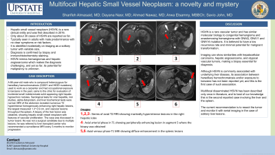
Figure: Figure 1: Series of axial T2 FS MRI showing markedly hyperintense lesions in the right hepatic lobe
Figure 2: Axial arterial phase in T1 showing peripherally enhancing lesion in segment 5 where the biopsy was obtained
Figure 3: Axial venous phase T1 FS MRI showing diffuse enhancement in the splenic lesions
Figure 2: Axial arterial phase in T1 showing peripherally enhancing lesion in segment 5 where the biopsy was obtained
Figure 3: Axial venous phase T1 FS MRI showing diffuse enhancement in the splenic lesions
Disclosures:
Sharifeh Almasaid indicated no relevant financial relationships.
Dayana Nasr indicated no relevant financial relationships.
Ahmad Nawaz indicated no relevant financial relationships.
Arwa Elsamny indicated no relevant financial relationships.
Savio John indicated no relevant financial relationships.
Sharifeh Almasaid, MD1, Dayana Nasr, MD2, Ahmad Nawaz, MD3, Arwa Elsamny, MBBCh3, Savio John, MD, FACG3. P3036 - Multifocal Hepatic Small Vessel Neoplasm: A Novelty and Mystery, ACG 2024 Annual Scientific Meeting Abstracts. Philadelphia, PA: American College of Gastroenterology.
