Monday Poster Session
Category: Liver
P3075 - Acute Liver Failure Leading to Undiagnosed Multiple Myeloma
Monday, October 28, 2024
10:30 AM - 4:00 PM ET
Location: Exhibit Hall E

Has Audio
- AP
Alessandra Pitco, MD
HCA Riverside Community Hospital
Riverside, CA
Presenting Author(s)
Regis Lee, DO, Alessandra Pitco, MD, Hoveda Mufti, MD, Waqqas Mirza, MD
HCA Riverside Community Hospital, Riverside, CA
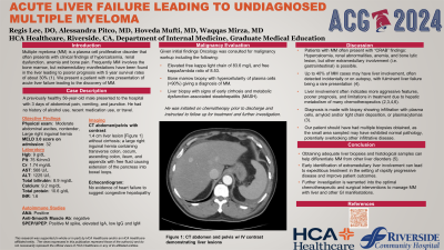
Introduction: Multiple myeloma (MM) is a plasma cell proliferative disorder that often presents with clinical findings of hypercalcemia, renal dysfunction, anemia and bone pain. Frequently MM involves the bone marrow, but extramedullary manifestations have been found in the liver leading to poorer prognosis. We present a patient initially presenting with acute liver failure leading to the discovery of MM.
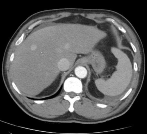
Case Description/Methods: A previously healthy 56-year-old male presented to the hospital with 3 days of abdominal pain, vomiting and jaundice. Vitals were stable. Physical exam showed ascites and a large right inguinal hernia. Lab work resulted with hemoglobin: 9 g/dL, platelets: 75 K/mm3, creatinine: 1.74 mg/dL, INR 1.6, AST: 568 U/L, ALT: 1225 U/L, total bilirubin: 8.9 mg/dL, and a negative hepatitis and infectious panel. CT abdomen and pelvis with intravenous contrast demonstrated a 1.4 cm liver lesion along with a large right inguinal hernia containing transverse colon, cecum, ascending colon, ilium, and appendix with free fluid causing extension of the pancreas into bowel loops. Although MELD 3.0 score was 32 on admission, there was no evidence of cirrhosis on MRI. Consideration for congestive hepatopathy was made, however echocardiogram showed no significant heart failure. Autoimmune studies revealed positive antinuclear antibody (ANA) and immunoglobulin levels leading to increased concern for autoimmune hepatitis (AIH). Further workup included serum protein and urine electrophoresis (SPEP and UPEP) showing an M spike along with an elevated serum IgA, low IgG and IgM. Given these findings Oncology was consulted for malignancy workup leading to an elevated free kappa light chain at 83.6 mg/L and free kappa/lambda ratio of 8.53. Ultimately, bone marrow biopsy demonstrated hypercellularity of plasma cells ( >90%) giving a diagnosis of MM. Liver biopsy was performed showing signs of early cirrhosis and metabolic dysfunction-associated steatohepatitis (MASH). He was initiated on chemotherapy prior to discharge and instructed to follow up for treatment and further investigation.
Discussion: Extramedullary involvement of the liver is rare with findings indicating poor prognosis due to limitations in treatment. Although the liver biopsy did not signify MM or AIH, additional biopsies should have been obtained which may have confirmed the diagnosis as up to 40% of MM cases have plasma cell infiltration of the liver. Acute liver failure should include a differential of MM as early detection can improve outcomes.

Disclosures:
Regis Lee, DO, Alessandra Pitco, MD, Hoveda Mufti, MD, Waqqas Mirza, MD. P3075 - Acute Liver Failure Leading to Undiagnosed Multiple Myeloma, ACG 2024 Annual Scientific Meeting Abstracts. Philadelphia, PA: American College of Gastroenterology.
HCA Riverside Community Hospital, Riverside, CA
Introduction: Multiple myeloma (MM) is a plasma cell proliferative disorder that often presents with clinical findings of hypercalcemia, renal dysfunction, anemia and bone pain. Frequently MM involves the bone marrow, but extramedullary manifestations have been found in the liver leading to poorer prognosis. We present a patient initially presenting with acute liver failure leading to the discovery of MM.
Case Description/Methods: A previously healthy 56-year-old male presented to the hospital with 3 days of abdominal pain, vomiting and jaundice. Vitals were stable. Physical exam showed ascites and a large right inguinal hernia. Lab work resulted with hemoglobin: 9 g/dL, platelets: 75 K/mm3, creatinine: 1.74 mg/dL, INR 1.6, AST: 568 U/L, ALT: 1225 U/L, total bilirubin: 8.9 mg/dL, and a negative hepatitis and infectious panel. CT abdomen and pelvis with intravenous contrast demonstrated a 1.4 cm liver lesion along with a large right inguinal hernia containing transverse colon, cecum, ascending colon, ilium, and appendix with free fluid causing extension of the pancreas into bowel loops. Although MELD 3.0 score was 32 on admission, there was no evidence of cirrhosis on MRI. Consideration for congestive hepatopathy was made, however echocardiogram showed no significant heart failure. Autoimmune studies revealed positive antinuclear antibody (ANA) and immunoglobulin levels leading to increased concern for autoimmune hepatitis (AIH). Further workup included serum protein and urine electrophoresis (SPEP and UPEP) showing an M spike along with an elevated serum IgA, low IgG and IgM. Given these findings Oncology was consulted for malignancy workup leading to an elevated free kappa light chain at 83.6 mg/L and free kappa/lambda ratio of 8.53. Ultimately, bone marrow biopsy demonstrated hypercellularity of plasma cells ( >90%) giving a diagnosis of MM. Liver biopsy was performed showing signs of early cirrhosis and metabolic dysfunction-associated steatohepatitis (MASH). He was initiated on chemotherapy prior to discharge and instructed to follow up for treatment and further investigation.
Discussion: Extramedullary involvement of the liver is rare with findings indicating poor prognosis due to limitations in treatment. Although the liver biopsy did not signify MM or AIH, additional biopsies should have been obtained which may have confirmed the diagnosis as up to 40% of MM cases have plasma cell infiltration of the liver. Acute liver failure should include a differential of MM as early detection can improve outcomes.

Figure: Liver lesions present on CT abdomen and pelvis with intravenous contrast
Disclosures:
Regis Lee indicated no relevant financial relationships.
Alessandra Pitco indicated no relevant financial relationships.
Hoveda Mufti indicated no relevant financial relationships.
Waqqas Mirza indicated no relevant financial relationships.
Regis Lee, DO, Alessandra Pitco, MD, Hoveda Mufti, MD, Waqqas Mirza, MD. P3075 - Acute Liver Failure Leading to Undiagnosed Multiple Myeloma, ACG 2024 Annual Scientific Meeting Abstracts. Philadelphia, PA: American College of Gastroenterology.
