Monday Poster Session
Category: Obesity
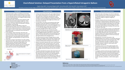
P3180 - Overinflated Solutions: Delayed Presentation From a Hyperinflated Intragastric Balloon
Monday, October 28, 2024
10:30 AM - 4:00 PM ET
Location: Exhibit Hall E

Has Audio

Raghav Bassi, MD
University of Central Florida College of Medicine
Gainesville, FL
Presenting Author(s)
Raghav Bassi, MD1, Emmanuel Magsino, MD, MS2, Sonia Alicea, MD2, Shin Zaw, DO2, Vinay Katukuri, MD2
1University of Central Florida College of Medicine, Gainesville, FL; 2University of Central Florida College of Medicine, Kissimmee, FL
Introduction: Endoscopic bariatric surgery is an effective and minimally invasive treatment option for patients looking to lose weight. The two endoscopic procedures approved by the US Food and Drug Administration are intragastric balloon (IGB) and gastric sleeve gastroplasty. Most IGBs are filled with 400-700cc of saline and methylene blue dye and occupy space in the stomach to increase satiety and delay gastric emptying.
Case Description/Methods: A 36-year-old Hispanic female, with a past medical history of class III obesity and status post placement of an Orbera IGB in Columbia two months prior to admission, presented with constant epigastric burning pain and bloody non-bilious emesis that progressively worsened over the past two weeks. She reported a total weight loss of 20 lbs since the placement of her IGB. On presentation, a computed tomography of the abdomen/pelvis with contrast revealed a 13.6 x 11.2 cm IGB with collapse of the distal stomach and associated esophageal dilation (Figure 1). The gastroenterology team was consulted and urgent endoscopic removal of the IGB was planned. She underwent an esophagogastroduodenoscopy which revealed the IGB situated in the proximal body of the stomach, covering nearly the entire luminal circumference, with mild erythema at the impingement site. The IGB was punctured using a 23-gauge injection needle, resulting in the leakage of saline mixed with methylene blue. A total of 850cc of fluid was aspirated, and the balloon was completely deflated before removal. Despite unsuccessful attempts using jumbo forceps, rat-tooth forceps, and a 3-prong grasper, the snare was eventually employed to capture one end of the balloon, and pull it through the esophagus with complete removal. Mineral oil was sprayed on the esophagus during the procedure to aid in the successful retrieval. The patient tolerated the procedure well and was discharged home the following day.
Discussion: Complications of IGBs include spontaneous hyperinflation from the contamination of IGB fluid with gas-forming microorganisms. Other causes of hyperinflation include overfilling during implantation resulting in the acute onset of obstructive symptoms within days. Our case above is unique in that the patient did not develop symptoms until months later. Studies have shown that the stomach volume decreases after weight loss, and it is postulated that as the IGB occupies more space in the gastric lumen, patients can develop delayed symptoms.

Disclosures:
Raghav Bassi, MD1, Emmanuel Magsino, MD, MS2, Sonia Alicea, MD2, Shin Zaw, DO2, Vinay Katukuri, MD2. P3180 - Overinflated Solutions: Delayed Presentation From a Hyperinflated Intragastric Balloon, ACG 2024 Annual Scientific Meeting Abstracts. Philadelphia, PA: American College of Gastroenterology.
1University of Central Florida College of Medicine, Gainesville, FL; 2University of Central Florida College of Medicine, Kissimmee, FL
Introduction: Endoscopic bariatric surgery is an effective and minimally invasive treatment option for patients looking to lose weight. The two endoscopic procedures approved by the US Food and Drug Administration are intragastric balloon (IGB) and gastric sleeve gastroplasty. Most IGBs are filled with 400-700cc of saline and methylene blue dye and occupy space in the stomach to increase satiety and delay gastric emptying.
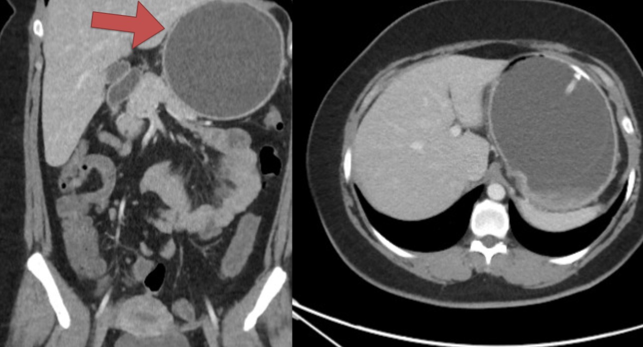
Case Description/Methods: A 36-year-old Hispanic female, with a past medical history of class III obesity and status post placement of an Orbera IGB in Columbia two months prior to admission, presented with constant epigastric burning pain and bloody non-bilious emesis that progressively worsened over the past two weeks. She reported a total weight loss of 20 lbs since the placement of her IGB. On presentation, a computed tomography of the abdomen/pelvis with contrast revealed a 13.6 x 11.2 cm IGB with collapse of the distal stomach and associated esophageal dilation (Figure 1). The gastroenterology team was consulted and urgent endoscopic removal of the IGB was planned. She underwent an esophagogastroduodenoscopy which revealed the IGB situated in the proximal body of the stomach, covering nearly the entire luminal circumference, with mild erythema at the impingement site. The IGB was punctured using a 23-gauge injection needle, resulting in the leakage of saline mixed with methylene blue. A total of 850cc of fluid was aspirated, and the balloon was completely deflated before removal. Despite unsuccessful attempts using jumbo forceps, rat-tooth forceps, and a 3-prong grasper, the snare was eventually employed to capture one end of the balloon, and pull it through the esophagus with complete removal. Mineral oil was sprayed on the esophagus during the procedure to aid in the successful retrieval. The patient tolerated the procedure well and was discharged home the following day.
Discussion: Complications of IGBs include spontaneous hyperinflation from the contamination of IGB fluid with gas-forming microorganisms. Other causes of hyperinflation include overfilling during implantation resulting in the acute onset of obstructive symptoms within days. Our case above is unique in that the patient did not develop symptoms until months later. Studies have shown that the stomach volume decreases after weight loss, and it is postulated that as the IGB occupies more space in the gastric lumen, patients can develop delayed symptoms.

Figure: Figure 1: CT of the abdomen and pelvis with contrast revealing a 13.6 x 11.2 cm IGB with collapse of the distal stomach and associated esophageal dilation.
Disclosures:
Raghav Bassi indicated no relevant financial relationships.
Emmanuel Magsino indicated no relevant financial relationships.
Sonia Alicea indicated no relevant financial relationships.
Shin Zaw indicated no relevant financial relationships.
Vinay Katukuri indicated no relevant financial relationships.
Raghav Bassi, MD1, Emmanuel Magsino, MD, MS2, Sonia Alicea, MD2, Shin Zaw, DO2, Vinay Katukuri, MD2. P3180 - Overinflated Solutions: Delayed Presentation From a Hyperinflated Intragastric Balloon, ACG 2024 Annual Scientific Meeting Abstracts. Philadelphia, PA: American College of Gastroenterology.
