Monday Poster Session
Category: Small Intestine
P3235 - An Atypical and Rare Presentation of Hodgkin Lymphoma Involving the Small Intestine
Monday, October 28, 2024
10:30 AM - 4:00 PM ET
Location: Exhibit Hall E

Has Audio

Kaitlyn Cronk, MD
University of Mississippi Medical Center
Jackson, MS
Presenting Author(s)
Kaitlyn Cronk, MD, Namrata Paladugu, BS, MD, Ismail Ganim, MD, Joydeep Chakraborty, MD
University of Mississippi Medical Center, Jackson, MS
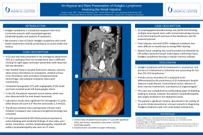
Introduction: Hodgkin lymphoma is a lymphoid neoplasm that most commonly presents with supradiaphragmatic lymphadenopathy and systemic B-symptoms. We present a case of Classic Hodgkin Lymphoma with small bowel involvement initially presenting as an acute stroke and melena.
Case Description/Methods: A 62-year-old male presented to the emergency department (ED) as a code grey due to difficulty moving his right upper and lower extremities with observed left eye deviation. Past medical history included Parkinson’s disease, seizures, deep venous thrombosis on enoxaparin, cerebral venous sinus thrombosis with secondary intraparenchymal hemorrhage, and subdural empyema status post craniectomy. Computed tomography (CT) with angiography of the head and neck revealed acute left basal ganglia infarct. In the ED, the patient reported recent melena which was also observed with his next bowel movement. Laboratory results were significant for hemoglobin 4.2 g/dL, white blood cell count 6.9 TH/cmm and lactate 2.3 mmol/L. Transfusion protocol and a pantoprazole infusion were initiated. Enoxaparin was reversed and gastroenterology was consulted. CT with gastrointestinal (GI) bleed protocol reported no active bleeding with an incidental finding of new celiac, peri-portal, mesenteric, and iliac lymphadenopathy. Isolated left axillary lymphadenopathy was seen on CT chest. An esophagogastroduodenoscopy was performed showing multiple clean-based ulcers with normal intervening mucosa in the third and fourth portions of the duodenum and the proximal jejunum. Ulcer biopsies returned CD30+ malignant neoplasm, but were difficult to classify due to strong PAX5 staining. Repeat tissue sampling was recommended and obtained via left axillary excisional lymph node biopsy confirming Classic Hodgkin Lymphoma favoring a nodular sclerosis subtype.
Discussion: Hodgkin lymphoma with GI involvement is uncommon, and primary GI involvement is extremely rare accounting for less than 5% of GI lymphomas. Primary versus secondary HL is proposed to be differentiated by the prominence of GI involvement despite adjacent nodal disease, normal blood counts, absence of bone marrow involvement, and absence of organomegaly. This case was complicated by confounding upper GI bleeding leading to anemia, however the patient’s lymphadenopathy was largely localized to the abdominal cavity. This patient’s significant melena, discovered in the setting of an acute stroke presented an unusual scenario in diagnosing Hodgkin lymphoma with rare small bowel involvement.

Disclosures:
Kaitlyn Cronk, MD, Namrata Paladugu, BS, MD, Ismail Ganim, MD, Joydeep Chakraborty, MD. P3235 - An Atypical and Rare Presentation of Hodgkin Lymphoma Involving the Small Intestine, ACG 2024 Annual Scientific Meeting Abstracts. Philadelphia, PA: American College of Gastroenterology.
University of Mississippi Medical Center, Jackson, MS
Introduction: Hodgkin lymphoma is a lymphoid neoplasm that most commonly presents with supradiaphragmatic lymphadenopathy and systemic B-symptoms. We present a case of Classic Hodgkin Lymphoma with small bowel involvement initially presenting as an acute stroke and melena.
Case Description/Methods: A 62-year-old male presented to the emergency department (ED) as a code grey due to difficulty moving his right upper and lower extremities with observed left eye deviation. Past medical history included Parkinson’s disease, seizures, deep venous thrombosis on enoxaparin, cerebral venous sinus thrombosis with secondary intraparenchymal hemorrhage, and subdural empyema status post craniectomy. Computed tomography (CT) with angiography of the head and neck revealed acute left basal ganglia infarct. In the ED, the patient reported recent melena which was also observed with his next bowel movement. Laboratory results were significant for hemoglobin 4.2 g/dL, white blood cell count 6.9 TH/cmm and lactate 2.3 mmol/L. Transfusion protocol and a pantoprazole infusion were initiated. Enoxaparin was reversed and gastroenterology was consulted. CT with gastrointestinal (GI) bleed protocol reported no active bleeding with an incidental finding of new celiac, peri-portal, mesenteric, and iliac lymphadenopathy. Isolated left axillary lymphadenopathy was seen on CT chest. An esophagogastroduodenoscopy was performed showing multiple clean-based ulcers with normal intervening mucosa in the third and fourth portions of the duodenum and the proximal jejunum. Ulcer biopsies returned CD30+ malignant neoplasm, but were difficult to classify due to strong PAX5 staining. Repeat tissue sampling was recommended and obtained via left axillary excisional lymph node biopsy confirming Classic Hodgkin Lymphoma favoring a nodular sclerosis subtype.
Discussion: Hodgkin lymphoma with GI involvement is uncommon, and primary GI involvement is extremely rare accounting for less than 5% of GI lymphomas. Primary versus secondary HL is proposed to be differentiated by the prominence of GI involvement despite adjacent nodal disease, normal blood counts, absence of bone marrow involvement, and absence of organomegaly. This case was complicated by confounding upper GI bleeding leading to anemia, however the patient’s lymphadenopathy was largely localized to the abdominal cavity. This patient’s significant melena, discovered in the setting of an acute stroke presented an unusual scenario in diagnosing Hodgkin lymphoma with rare small bowel involvement.

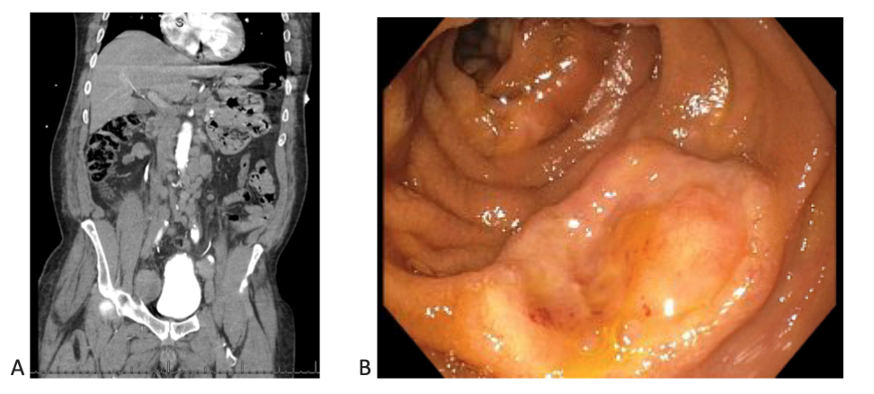
Figure: A. Coronal view of abdominal and pelvic CT scan with significant celiac, peri-portal, mesenteric, para-aortic, and iliac lymphadenopathy.
B. Duodenal ulcer visualized on upper endoscopy.
B. Duodenal ulcer visualized on upper endoscopy.
Disclosures:
Kaitlyn Cronk indicated no relevant financial relationships.
Namrata Paladugu indicated no relevant financial relationships.
Ismail Ganim indicated no relevant financial relationships.
Joydeep Chakraborty indicated no relevant financial relationships.
Kaitlyn Cronk, MD, Namrata Paladugu, BS, MD, Ismail Ganim, MD, Joydeep Chakraborty, MD. P3235 - An Atypical and Rare Presentation of Hodgkin Lymphoma Involving the Small Intestine, ACG 2024 Annual Scientific Meeting Abstracts. Philadelphia, PA: American College of Gastroenterology.
