Monday Poster Session
Category: Small Intestine
P3237 - A Rare Case of Duodenal Diverticulitis: RUQ Pain That Isn’t Acute Cholecystitis?
Monday, October 28, 2024
10:30 AM - 4:00 PM ET
Location: Exhibit Hall E

Has Audio
- RS
Robinderpal Sandhu, DO
Mount Sinai Morningside, Icahn School of Medicine at Mount Sinai
New York, NY
Presenting Author(s)
Robinderpal Sandhu, DO1, Adam Tillowitz, MD2, Frank Nelson, MD3
1Mount Sinai Morningside, Icahn School of Medicine at Mount Sinai, New York, NY; 2Icahn School of Medicine at Mount Sinai Morningside/West, New York, NY; 3Mount Sinai West, Icahn School of Medicine at Mount Sinai, New York, NY
Introduction: The differential for right upper quadrant (RUQ) pain includes liver and gallbladder disorders, inflammatory bowel disease, pancreatic disorders, pulmonary disease, malignancy, and rarely diverticular disease. Duodenal diverticulum is a common incidental finding on computed tomography (CT) scan however, only about 10% cause symptoms with less than 1% requiring treatment due to perforation, hemorrhage, or obstruction1. Here we present a rare care of perforated acute duodenal diverticulitis in a 77-year-old female who presented with right upper quadrant pain.
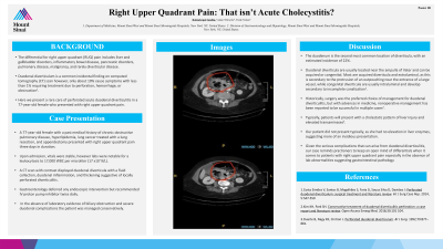
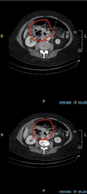
Case Description/Methods: A 77-year-old female with a past medical history of chronic obstructive pulmonary disease, hyperlipidemia, lung cancer treated with a lung resection, and appendectomy presented with right upper quadrant pain three days in duration. Upon admission, vitals were stable, however labs were notable for a leukocytosis to 17,000 WBC per microliter (17 x10ˆ9/L). A CT scan with contrast displayed duodenal diverticula with a fluid collection, duodenal inflammation, and thickening suggestive of locally perforated diverticulitis. Gastroenterology deferred any endoscopic intervention but recommended IV proton pump inhibitor twice daily. In the absence of laboratory evidence of biliary obstruction and severe duodenal complications the patient was managed conservatively.
Discussion: The duodenum is the second most common location of diverticula, with an estimated incidence of 22%. Duodenal diverticula are usually located near the ampulla of Vater and can be acquired or congenital. Most are acquired diverticula and extraluminal, as this is secondary to the protrusion of an outpouching near the entrance of a large vessel, while congenital diverticula are usually intraluminal and develop secondary to incomplete canalization1. Historically, surgery was the preferred choice of management for duodenal diverticulitis, but with advances in medicine, nonoperative management has been reported to be successful in multiple cases2. Typically, patients will present with a cholestatic pattern of liver injury and elevated transaminases3. Our patient did not present typically, as she had no elevation in liver enzymes, suggesting more of an insidious presentation. Given the serious complications that can arise from duodenal diverticulitis, our case reminds practioners to keep an open mind of differentials when it comes to patients with right upper quadrant pain especially in the absence of lab abnormalities suggesting gastrointestinal pathology.

Disclosures:
Robinderpal Sandhu, DO1, Adam Tillowitz, MD2, Frank Nelson, MD3. P3237 - A Rare Case of Duodenal Diverticulitis: RUQ Pain That Isn’t Acute Cholecystitis?, ACG 2024 Annual Scientific Meeting Abstracts. Philadelphia, PA: American College of Gastroenterology.
1Mount Sinai Morningside, Icahn School of Medicine at Mount Sinai, New York, NY; 2Icahn School of Medicine at Mount Sinai Morningside/West, New York, NY; 3Mount Sinai West, Icahn School of Medicine at Mount Sinai, New York, NY
Introduction: The differential for right upper quadrant (RUQ) pain includes liver and gallbladder disorders, inflammatory bowel disease, pancreatic disorders, pulmonary disease, malignancy, and rarely diverticular disease. Duodenal diverticulum is a common incidental finding on computed tomography (CT) scan however, only about 10% cause symptoms with less than 1% requiring treatment due to perforation, hemorrhage, or obstruction1. Here we present a rare care of perforated acute duodenal diverticulitis in a 77-year-old female who presented with right upper quadrant pain.
Case Description/Methods: A 77-year-old female with a past medical history of chronic obstructive pulmonary disease, hyperlipidemia, lung cancer treated with a lung resection, and appendectomy presented with right upper quadrant pain three days in duration. Upon admission, vitals were stable, however labs were notable for a leukocytosis to 17,000 WBC per microliter (17 x10ˆ9/L). A CT scan with contrast displayed duodenal diverticula with a fluid collection, duodenal inflammation, and thickening suggestive of locally perforated diverticulitis. Gastroenterology deferred any endoscopic intervention but recommended IV proton pump inhibitor twice daily. In the absence of laboratory evidence of biliary obstruction and severe duodenal complications the patient was managed conservatively.
Discussion: The duodenum is the second most common location of diverticula, with an estimated incidence of 22%. Duodenal diverticula are usually located near the ampulla of Vater and can be acquired or congenital. Most are acquired diverticula and extraluminal, as this is secondary to the protrusion of an outpouching near the entrance of a large vessel, while congenital diverticula are usually intraluminal and develop secondary to incomplete canalization1. Historically, surgery was the preferred choice of management for duodenal diverticulitis, but with advances in medicine, nonoperative management has been reported to be successful in multiple cases2. Typically, patients will present with a cholestatic pattern of liver injury and elevated transaminases3. Our patient did not present typically, as she had no elevation in liver enzymes, suggesting more of an insidious presentation. Given the serious complications that can arise from duodenal diverticulitis, our case reminds practioners to keep an open mind of differentials when it comes to patients with right upper quadrant pain especially in the absence of lab abnormalities suggesting gastrointestinal pathology.

Figure: A. Perforated duodenal diverticulum encircled in red
Disclosures:
Robinderpal Sandhu indicated no relevant financial relationships.
Adam Tillowitz indicated no relevant financial relationships.
Frank Nelson indicated no relevant financial relationships.
Robinderpal Sandhu, DO1, Adam Tillowitz, MD2, Frank Nelson, MD3. P3237 - A Rare Case of Duodenal Diverticulitis: RUQ Pain That Isn’t Acute Cholecystitis?, ACG 2024 Annual Scientific Meeting Abstracts. Philadelphia, PA: American College of Gastroenterology.
