Monday Poster Session
Category: Small Intestine
P3244 - A Case of Elusive Small Bowel Melanoma
Monday, October 28, 2024
10:30 AM - 4:00 PM ET
Location: Exhibit Hall E

Has Audio
- KR
Kristen Ruckstuhl, DO, MS
Case Western Reserve University / MetroHealth
Cleveland, OH
Presenting Author(s)
Kristen Ruckstuhl, DO, MS, Sara Kamionkowski, DO, Nisheet Waghray, MD
Case Western Reserve University / MetroHealth, Cleveland, OH
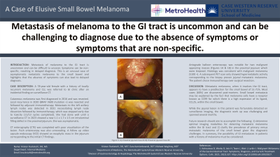
Introduction: Metastasis of melanoma to the GI tract is uncommon and can be difficult to uncover. Symptoms can be non-specific, resulting in delayed diagnosis. This is an unusual case of asymptomatic metastatic melanoma to the small bowel and highlights that the absence of symptoms can also lead to delayed diagnosis.
Case Description/Methods: A 78-year-old female with a history of locally recurrent melanoma and CLL was referred to GI clinic after an incidental finding on surveillance CT. Cutaneous melanoma was first detected in 2016, which was resected. Local recurrence in 2020 (BRAF V600 mutation +) was resected and followed by adjuvant immunotherapy. Metastasis to the left axillary lymph nodes was detected in 2021 necessitating lymph node dissection followed by immunotherapy, which was stopped early due to toxicity (11/13 cycles completed). She had done well until a surveillance CT in 2023 showed a new 1.1 x 1.7 x 1.5 cm intraluminal filling defect in the proximal jejunum. She was asymptomatic. CT enterography (CTE) was completed with poor visualization of the lesion. Push enteroscopy was also unrevealing. Follow up video capsule endoscopy (VCE) showed an exophytic mass in the jejunum corresponding to the initial CT findings. Antegrade balloon enteroscopy was notable for two malignant appearing masses (Figures 1a &1b) in the proximal jejunum which were biopsied. Histology was consistent with malignant melanoma (S100 +). A subsequent PET scan only showed hypermetabolic activity corresponding to the biopsy proven jejunal metastatic melanoma. The patient chose immunotherapy over surgical resection.
Discussion: Metastatic melanoma, when it involves the GI tract, appears to have a predilection for the small bowel of 51-71%. Most cases (60%) are discovered post-mortem. Small bowel metastasis may be explained by the fact that melanoma cells have a receptor known as CCR9 for which there is a high expression of its ligand, CCL25, within the small bowel. While the jejunal lesion in this patient was fortunately detected on surveillance imaging, the diagnostic work up was challenging and spanned several months. Future research should aim to accomplish the following: 1) determine optimal imaging modalities for detecting metastatic melanoma within the GI tract and 2) clarify the prevalence of primary versus metastatic melanoma of the small bowel given the diagnostic challenges. In summary, the possibility of GI metastases in patients with a history of melanoma must be considered.

Disclosures:
Kristen Ruckstuhl, DO, MS, Sara Kamionkowski, DO, Nisheet Waghray, MD. P3244 - A Case of Elusive Small Bowel Melanoma, ACG 2024 Annual Scientific Meeting Abstracts. Philadelphia, PA: American College of Gastroenterology.
Case Western Reserve University / MetroHealth, Cleveland, OH
Introduction: Metastasis of melanoma to the GI tract is uncommon and can be difficult to uncover. Symptoms can be non-specific, resulting in delayed diagnosis. This is an unusual case of asymptomatic metastatic melanoma to the small bowel and highlights that the absence of symptoms can also lead to delayed diagnosis.
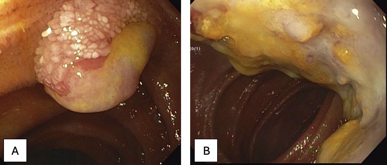
Case Description/Methods: A 78-year-old female with a history of locally recurrent melanoma and CLL was referred to GI clinic after an incidental finding on surveillance CT. Cutaneous melanoma was first detected in 2016, which was resected. Local recurrence in 2020 (BRAF V600 mutation +) was resected and followed by adjuvant immunotherapy. Metastasis to the left axillary lymph nodes was detected in 2021 necessitating lymph node dissection followed by immunotherapy, which was stopped early due to toxicity (11/13 cycles completed). She had done well until a surveillance CT in 2023 showed a new 1.1 x 1.7 x 1.5 cm intraluminal filling defect in the proximal jejunum. She was asymptomatic. CT enterography (CTE) was completed with poor visualization of the lesion. Push enteroscopy was also unrevealing. Follow up video capsule endoscopy (VCE) showed an exophytic mass in the jejunum corresponding to the initial CT findings. Antegrade balloon enteroscopy was notable for two malignant appearing masses (Figures 1a &1b) in the proximal jejunum which were biopsied. Histology was consistent with malignant melanoma (S100 +). A subsequent PET scan only showed hypermetabolic activity corresponding to the biopsy proven jejunal metastatic melanoma. The patient chose immunotherapy over surgical resection.
Discussion: Metastatic melanoma, when it involves the GI tract, appears to have a predilection for the small bowel of 51-71%. Most cases (60%) are discovered post-mortem. Small bowel metastasis may be explained by the fact that melanoma cells have a receptor known as CCR9 for which there is a high expression of its ligand, CCL25, within the small bowel. While the jejunal lesion in this patient was fortunately detected on surveillance imaging, the diagnostic work up was challenging and spanned several months. Future research should aim to accomplish the following: 1) determine optimal imaging modalities for detecting metastatic melanoma within the GI tract and 2) clarify the prevalence of primary versus metastatic melanoma of the small bowel given the diagnostic challenges. In summary, the possibility of GI metastases in patients with a history of melanoma must be considered.

Figure: Figure 1: A) Proximal jejunal mass #1 B) Proximal jejunal mass #2
Disclosures:
Kristen Ruckstuhl indicated no relevant financial relationships.
Sara Kamionkowski indicated no relevant financial relationships.
Nisheet Waghray indicated no relevant financial relationships.
Kristen Ruckstuhl, DO, MS, Sara Kamionkowski, DO, Nisheet Waghray, MD. P3244 - A Case of Elusive Small Bowel Melanoma, ACG 2024 Annual Scientific Meeting Abstracts. Philadelphia, PA: American College of Gastroenterology.
