Monday Poster Session
Category: Small Intestine
P3260 - Navigating Rare Presentations: Recurrent Burkitt Lymphoma Presenting as a Jejunal Mass
Monday, October 28, 2024
10:30 AM - 4:00 PM ET
Location: Exhibit Hall E

Has Audio

Tejas Nikumbh, MD
The Wright Center for Graduate Medical Education
Scranton, PA
Presenting Author(s)
Tejas Nikumbh, MD1, Archit Garg, MD2, Tushar Abhinav, MD3, Nivesh Yadav, MD3, Udit Asija, MD3, Amit Sohagia, MD4
1The Wright Center for Graduate Medical Education, Scanton, PA; 2Saint Peter's University Hospital, New Brunswick, NJ; 3The Wright Center for Graduate Medical Education, Scranton, PA; 4Geisinger Community Medical Center, Scranton, PA
Introduction: Burkitt lymphoma (BL) is a highly aggressive tumor constituting 1-5% of non- Hodgkin’s B cell lymphomas. BL when present in the gastrointestinal tract, usually involves the stomach. BL originating in intestines account for about 9% cases, mostly affecting the ileocecal region, presentation of BL in the jejunum is relatively rare.
Case Description/Methods: A 37-year-old male presented with abdominal pain for 5 days. He reported the pain as localized to the lower abdomen, with a severity of 6/10, accompanied by nausea and cramping.
In 2010, he underwent right radical orchiectomy for stage III non-seminomatous testicular carcinoma, with pathology indicating 90% embryonal and 10% yolk sac components with vascular invasion. He completed 5 cycles of chemotherapy with etoposide and platinum. In 2015, he was diagnosed with Burkitt lymphoma after a PET-CT scan revealed an enlarged gastrohepatic ligament lymph node. He received 6 cycles of hyper-CVAD therapy.
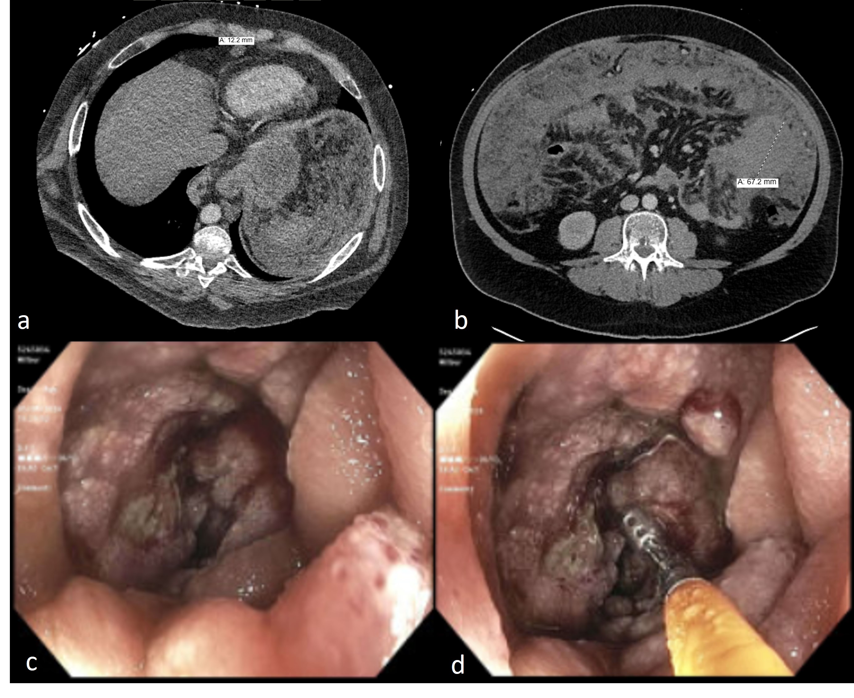
Initial lab results showed acute kidney injury with lactic acidosis. CT scan of the abdomen revealed small bowel loop wall thickening in left hemiabdomen, measuring up to 6.7 cm, with peritoneal carcinomatosis (fig a,b). Incidentally, segmental and subsegmental pulmonary emboli (PE) in the right lower lobe was also noted, and heparin drip was started. He was treated with rasburicase due to concerns of spontaneous tumor lysis syndrome.
IR-guided biopsy of the peritoneal carcinomatosis was considered, but an endoscopic biopsy of the jejunal mass was preferred for a definitive pathological diagnosis. The patient underwent push enteroscopy for a biopsy of the jejunal mass (fig 1 c,d), which returned positive for CD45, CD20, BCL6, and c-Myc, and negative for CK AE 1/3, BCL-2, SCL L4, and OCD 4, suggesting recurrence of Burkitt lymphoma.
Due to increased oxygen requirements and worsening lactic acidosis, the patient was transferred to the ICU for further management. He required intubation for respiratory failure and was placed on multiple pressors. Patient suffered multiple cardiac arrests, and despite best efforts, the patient expired.
Discussion: This case underscores the complexity and aggressive nature of BL, particularly when it presents in rare locations such as the jejunum. Early recognition and prompt intervention are critical in managing such aggressive tumors, though the prognosis can remain poor due to rapid progression and complications like PE, as seen in this patient's unfortunate outcome.

Disclosures:
Tejas Nikumbh, MD1, Archit Garg, MD2, Tushar Abhinav, MD3, Nivesh Yadav, MD3, Udit Asija, MD3, Amit Sohagia, MD4. P3260 - Navigating Rare Presentations: Recurrent Burkitt Lymphoma Presenting as a Jejunal Mass, ACG 2024 Annual Scientific Meeting Abstracts. Philadelphia, PA: American College of Gastroenterology.
1The Wright Center for Graduate Medical Education, Scanton, PA; 2Saint Peter's University Hospital, New Brunswick, NJ; 3The Wright Center for Graduate Medical Education, Scranton, PA; 4Geisinger Community Medical Center, Scranton, PA
Introduction: Burkitt lymphoma (BL) is a highly aggressive tumor constituting 1-5% of non- Hodgkin’s B cell lymphomas. BL when present in the gastrointestinal tract, usually involves the stomach. BL originating in intestines account for about 9% cases, mostly affecting the ileocecal region, presentation of BL in the jejunum is relatively rare.
Case Description/Methods: A 37-year-old male presented with abdominal pain for 5 days. He reported the pain as localized to the lower abdomen, with a severity of 6/10, accompanied by nausea and cramping.
In 2010, he underwent right radical orchiectomy for stage III non-seminomatous testicular carcinoma, with pathology indicating 90% embryonal and 10% yolk sac components with vascular invasion. He completed 5 cycles of chemotherapy with etoposide and platinum. In 2015, he was diagnosed with Burkitt lymphoma after a PET-CT scan revealed an enlarged gastrohepatic ligament lymph node. He received 6 cycles of hyper-CVAD therapy.
Initial lab results showed acute kidney injury with lactic acidosis. CT scan of the abdomen revealed small bowel loop wall thickening in left hemiabdomen, measuring up to 6.7 cm, with peritoneal carcinomatosis (fig a,b). Incidentally, segmental and subsegmental pulmonary emboli (PE) in the right lower lobe was also noted, and heparin drip was started. He was treated with rasburicase due to concerns of spontaneous tumor lysis syndrome.
IR-guided biopsy of the peritoneal carcinomatosis was considered, but an endoscopic biopsy of the jejunal mass was preferred for a definitive pathological diagnosis. The patient underwent push enteroscopy for a biopsy of the jejunal mass (fig 1 c,d), which returned positive for CD45, CD20, BCL6, and c-Myc, and negative for CK AE 1/3, BCL-2, SCL L4, and OCD 4, suggesting recurrence of Burkitt lymphoma.
Due to increased oxygen requirements and worsening lactic acidosis, the patient was transferred to the ICU for further management. He required intubation for respiratory failure and was placed on multiple pressors. Patient suffered multiple cardiac arrests, and despite best efforts, the patient expired.
Discussion: This case underscores the complexity and aggressive nature of BL, particularly when it presents in rare locations such as the jejunum. Early recognition and prompt intervention are critical in managing such aggressive tumors, though the prognosis can remain poor due to rapid progression and complications like PE, as seen in this patient's unfortunate outcome.

Figure: Figure 1: CT abdomen reveals a. Multiple nodules suggestive of peritoneal carcinomatosis, b. 6.7 cm jejunal mass in left hemiabdomen; Push enteroscopy showed c. Ulcerated mass with no bleed in the jejunum. d. Biopsies were taken with cold forceps for histopathology.
Disclosures:
Tejas Nikumbh indicated no relevant financial relationships.
Archit Garg indicated no relevant financial relationships.
Tushar Abhinav indicated no relevant financial relationships.
Nivesh Yadav indicated no relevant financial relationships.
Udit Asija indicated no relevant financial relationships.
Amit Sohagia indicated no relevant financial relationships.
Tejas Nikumbh, MD1, Archit Garg, MD2, Tushar Abhinav, MD3, Nivesh Yadav, MD3, Udit Asija, MD3, Amit Sohagia, MD4. P3260 - Navigating Rare Presentations: Recurrent Burkitt Lymphoma Presenting as a Jejunal Mass, ACG 2024 Annual Scientific Meeting Abstracts. Philadelphia, PA: American College of Gastroenterology.
