Monday Poster Session
Category: Small Intestine
P3286 - Primary Ewing Sarcoma of the Duodenum
Monday, October 28, 2024
10:30 AM - 4:00 PM ET
Location: Exhibit Hall E

Has Audio

Deva Christine T. Tojong, MD
Medical College of Georgia at Augusta University
Augusta, GA
Presenting Author(s)
Deva Christine T. Tojong, MD1, Gomathy A. Nageswaran, MBBS2, Ronan Calibuso, MD3, John Erikson Yap, MD, MBA, FACG4, Alexander Nguyen, MD5
1Medical College of Georgia at Augusta University, Augusta, GA; 2University of Arkansas for Medical Sciences, Little Rock, AR; 3Jersey City, NJ; 4Metrodora Institute, West Valley City, UT; 5Southbay Gastroenterology Medical Group, Torrance, CA
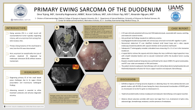
Introduction: Ewing sarcoma (ES) is a small round cell neuroectodermal tumor typically originating in bone, with an uncommon form arising from extra-osseous sites. Primary ES of the small bowel is rarer, with fewer than 50 cases documented in the literature. Here, we present an exceptionally rare case of primary duodenal ES diagnosed by endoscopic ultrasound (EUS) without osseous involvement.
Case Description/Methods: A 25-year-old male without medical problems presented to clinic with recurrent right upper quadrant abdominal pain, associated with nausea, vomiting, and melena for several months. His vitals and physical exam were normal. Lab findings revealed iron deficiency anemia. Esophagogastroduodenoscopy (EGD) and colonoscopy came back normal with negative H. pylori. A noncontrast abdominal CT scan identified clustered small bowel loops and a video capsule endoscopy showed duodenitis with capsule retention at the proximal small bowel. Subsequent CT enterography revealed a lobulated mass measuring 13 x 11 x 6 cm in the transverse duodenum. A repeat EGD to retrieve the capsule and EUS to biopsy the mass confirmed a large hypoechoic mass with smooth lobulated borders and no vascular involvement within the duodenal lumen causing obstruction. Biopsies revealed duodenal ES confirmed by the classic EWSR1-FL1 gene translocation. PET scan ruled out metastasis or FDG-avid lesions. The patient started neoadjuvant chemotherapy with vincristine/doxorubicin/cyclophosphamide and ifosfamide/etoposide with plans for future surgical resection and adjuvant chemotherapy.
Discussion: Diagnosing primary ES of the small bowel poses challenges due to its vague clinical presentation and inconclusive radiological findings. Definitive diagnosis depends on obtaining tissue for immunohistochemistry and genetic studies, with 85-90% of cases having the classic chromosomal translocation of ESWR1 gene on chromosome 22 to the FL1 gene on chromosome 11. Treatment predominantly involves neoadjuvant and adjuvant chemotherapy and surgical resection. Adverse clinical outcomes correlate with tumor size larger than 5 cm, involvement of pelvic sites, advanced age, chemotherapy resistance, and the presence of metastases. Advancing research is essential to refine treatment protocols and enhance diagnostic accuracy.

Disclosures:
Deva Christine T. Tojong, MD1, Gomathy A. Nageswaran, MBBS2, Ronan Calibuso, MD3, John Erikson Yap, MD, MBA, FACG4, Alexander Nguyen, MD5. P3286 - Primary Ewing Sarcoma of the Duodenum, ACG 2024 Annual Scientific Meeting Abstracts. Philadelphia, PA: American College of Gastroenterology.
1Medical College of Georgia at Augusta University, Augusta, GA; 2University of Arkansas for Medical Sciences, Little Rock, AR; 3Jersey City, NJ; 4Metrodora Institute, West Valley City, UT; 5Southbay Gastroenterology Medical Group, Torrance, CA
Introduction: Ewing sarcoma (ES) is a small round cell neuroectodermal tumor typically originating in bone, with an uncommon form arising from extra-osseous sites. Primary ES of the small bowel is rarer, with fewer than 50 cases documented in the literature. Here, we present an exceptionally rare case of primary duodenal ES diagnosed by endoscopic ultrasound (EUS) without osseous involvement.
Case Description/Methods: A 25-year-old male without medical problems presented to clinic with recurrent right upper quadrant abdominal pain, associated with nausea, vomiting, and melena for several months. His vitals and physical exam were normal. Lab findings revealed iron deficiency anemia. Esophagogastroduodenoscopy (EGD) and colonoscopy came back normal with negative H. pylori. A noncontrast abdominal CT scan identified clustered small bowel loops and a video capsule endoscopy showed duodenitis with capsule retention at the proximal small bowel. Subsequent CT enterography revealed a lobulated mass measuring 13 x 11 x 6 cm in the transverse duodenum. A repeat EGD to retrieve the capsule and EUS to biopsy the mass confirmed a large hypoechoic mass with smooth lobulated borders and no vascular involvement within the duodenal lumen causing obstruction. Biopsies revealed duodenal ES confirmed by the classic EWSR1-FL1 gene translocation. PET scan ruled out metastasis or FDG-avid lesions. The patient started neoadjuvant chemotherapy with vincristine/doxorubicin/cyclophosphamide and ifosfamide/etoposide with plans for future surgical resection and adjuvant chemotherapy.
Discussion: Diagnosing primary ES of the small bowel poses challenges due to its vague clinical presentation and inconclusive radiological findings. Definitive diagnosis depends on obtaining tissue for immunohistochemistry and genetic studies, with 85-90% of cases having the classic chromosomal translocation of ESWR1 gene on chromosome 22 to the FL1 gene on chromosome 11. Treatment predominantly involves neoadjuvant and adjuvant chemotherapy and surgical resection. Adverse clinical outcomes correlate with tumor size larger than 5 cm, involvement of pelvic sites, advanced age, chemotherapy resistance, and the presence of metastases. Advancing research is essential to refine treatment protocols and enhance diagnostic accuracy.

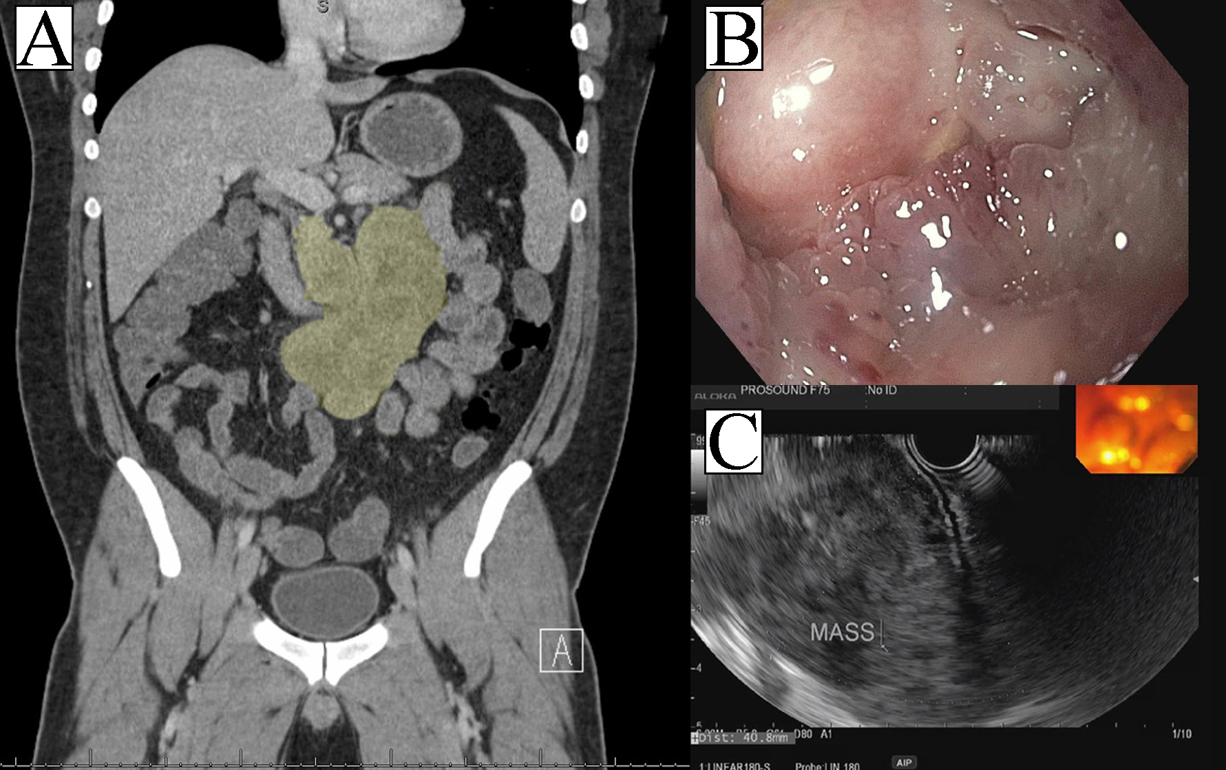
Figure: Figure A. CT abdomen with large lobulated mass (highlighted) located at the 3rd portion/transverse duodenum, measuring 13 x 11 x 6 cm.
Figure B. Endoscopic picture of 3rd portion of duodenum with wall edema and irregular mucosa indicative of a friable mass.
Figure C. EUS showed hypoechoic large mass with smooth lobulated borders and no vascularity.
Figure B. Endoscopic picture of 3rd portion of duodenum with wall edema and irregular mucosa indicative of a friable mass.
Figure C. EUS showed hypoechoic large mass with smooth lobulated borders and no vascularity.
Disclosures:
Deva Christine Tojong indicated no relevant financial relationships.
Gomathy Nageswaran indicated no relevant financial relationships.
Ronan Calibuso indicated no relevant financial relationships.
John Erikson Yap indicated no relevant financial relationships.
Alexander Nguyen indicated no relevant financial relationships.
Deva Christine T. Tojong, MD1, Gomathy A. Nageswaran, MBBS2, Ronan Calibuso, MD3, John Erikson Yap, MD, MBA, FACG4, Alexander Nguyen, MD5. P3286 - Primary Ewing Sarcoma of the Duodenum, ACG 2024 Annual Scientific Meeting Abstracts. Philadelphia, PA: American College of Gastroenterology.
