Monday Poster Session
Category: Small Intestine
P3299 - Large Brunner’s Gland Hamartoma as a Rare Culprit of Upper GI Bleed
Monday, October 28, 2024
10:30 AM - 4:00 PM ET
Location: Exhibit Hall E

Has Audio

Samantha Rubin, DO
Lenox Hill Hospital, Northwell Health
New York, NY
Presenting Author(s)
Samantha Rubin, DO1, Neil Gambhir, DO1, Noah Y. Mahpour, MD1, Michael Ma, MD2
1Lenox Hill Hospital, Northwell Health, New York, NY; 2Northwell Health, New York, NY
Introduction: Brunner’s Gland Hamartoma (BGH) is a benign tumor of the duodenum with diameter varying from 1.0 to 2.0 cm, rarely larger than 5.0 cm. It is uncommon, with a frequency of less than 1.0% among the primary tumors of the small intestine. Among patients who undergo routine esophagogastroduodenoscopy (EGD), BGH can be found in 0.01–0.07%. Most patients are asymptomatic, however rare cases present with gastrointestinal bleeding. Here, we report a rare case of upper gastrointestinal (GI) bleed due to a pedunculated BGH with ulceration measuring 5 cm.
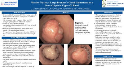
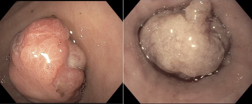
Case Description/Methods: An 88 year-old female with past medical history of Barrett's esophagus and recent provoked DVT on Eliquis, presented to the emergency room with melena. She was hemodynamically stable. Her laboratory values were significant for hemoglobin of 6.5, for which she received a unit of blood. She underwent endoscopy which identified esophagitis, fundic gland type polyps, and a large ulcerated pedunculated polyp traversing the gastric pylorus, seen in figure 1. The polyp was removed endoscopically with hot snare polypectomy. She had no further melena during admission and remained stable. Pathology returned as Brunner’s gland hamartoma measuring 5cm. She was discharged with close outpatient GI follow-up.
Discussion: This case reports a rare occurrence of a large bleeding BGH found crossing the gastric pylorus, noteworthy due to its potential to cause significant symptoms like melena, and only 4% of cases reported in this location. The pathogenesis of BGH is not well understood but is thought to involve hyperplasia of Brunner's glands due to chronic inflammation. Often asymptomatic and incidentally discovered during endoscopic or imaging studies, BGH can present with abdominal pain and gastrointestinal bleeding when symptomatic. Endoscopic examination typically reveals a submucosal mass with a smooth, polypoid appearance, however our case presented with an ulcerated polypoid lesion on a stalk. Biopsy and histopathological examination are essential to confirm the diagnosis and exclude malignancy. This case underscores the importance of considering BGH in the differential diagnosis of gastrointestinal bleeding, particularly melena. The rare trans-pyloric location of the hamartoma highlights the need for thorough diagnostic approaches and increased clinician awareness of this rare entity as a cause of upper GI bleed.

Disclosures:
Samantha Rubin, DO1, Neil Gambhir, DO1, Noah Y. Mahpour, MD1, Michael Ma, MD2. P3299 - Large Brunner’s Gland Hamartoma as a Rare Culprit of Upper GI Bleed, ACG 2024 Annual Scientific Meeting Abstracts. Philadelphia, PA: American College of Gastroenterology.
1Lenox Hill Hospital, Northwell Health, New York, NY; 2Northwell Health, New York, NY
Introduction: Brunner’s Gland Hamartoma (BGH) is a benign tumor of the duodenum with diameter varying from 1.0 to 2.0 cm, rarely larger than 5.0 cm. It is uncommon, with a frequency of less than 1.0% among the primary tumors of the small intestine. Among patients who undergo routine esophagogastroduodenoscopy (EGD), BGH can be found in 0.01–0.07%. Most patients are asymptomatic, however rare cases present with gastrointestinal bleeding. Here, we report a rare case of upper gastrointestinal (GI) bleed due to a pedunculated BGH with ulceration measuring 5 cm.
Case Description/Methods: An 88 year-old female with past medical history of Barrett's esophagus and recent provoked DVT on Eliquis, presented to the emergency room with melena. She was hemodynamically stable. Her laboratory values were significant for hemoglobin of 6.5, for which she received a unit of blood. She underwent endoscopy which identified esophagitis, fundic gland type polyps, and a large ulcerated pedunculated polyp traversing the gastric pylorus, seen in figure 1. The polyp was removed endoscopically with hot snare polypectomy. She had no further melena during admission and remained stable. Pathology returned as Brunner’s gland hamartoma measuring 5cm. She was discharged with close outpatient GI follow-up.
Discussion: This case reports a rare occurrence of a large bleeding BGH found crossing the gastric pylorus, noteworthy due to its potential to cause significant symptoms like melena, and only 4% of cases reported in this location. The pathogenesis of BGH is not well understood but is thought to involve hyperplasia of Brunner's glands due to chronic inflammation. Often asymptomatic and incidentally discovered during endoscopic or imaging studies, BGH can present with abdominal pain and gastrointestinal bleeding when symptomatic. Endoscopic examination typically reveals a submucosal mass with a smooth, polypoid appearance, however our case presented with an ulcerated polypoid lesion on a stalk. Biopsy and histopathological examination are essential to confirm the diagnosis and exclude malignancy. This case underscores the importance of considering BGH in the differential diagnosis of gastrointestinal bleeding, particularly melena. The rare trans-pyloric location of the hamartoma highlights the need for thorough diagnostic approaches and increased clinician awareness of this rare entity as a cause of upper GI bleed.

Figure: Brunner's Gland Hamartoma seen traversing the gastric pylorus
Disclosures:
Samantha Rubin indicated no relevant financial relationships.
Neil Gambhir indicated no relevant financial relationships.
Noah Mahpour indicated no relevant financial relationships.
Michael Ma indicated no relevant financial relationships.
Samantha Rubin, DO1, Neil Gambhir, DO1, Noah Y. Mahpour, MD1, Michael Ma, MD2. P3299 - Large Brunner’s Gland Hamartoma as a Rare Culprit of Upper GI Bleed, ACG 2024 Annual Scientific Meeting Abstracts. Philadelphia, PA: American College of Gastroenterology.
