Monday Poster Session
Category: Small Intestine
P3309 - Rare Case of Metastatic Renal Cell Carcinoma to the Duodenum
Monday, October 28, 2024
10:30 AM - 4:00 PM ET
Location: Exhibit Hall E

Has Audio
- SR
Sana Rabeeah, MD
University of Toledo
Toledo, OH
Presenting Author(s)
Sana Rabeeah, MD1, Sahithi Chinnam, DO1, Ahmed El Rahyel, MD1, Ali Wakil, MD2, Hasan Al-Obaidi, MD3, Omar Saab, MD4, Sami Ghazaleh, MD5, Yaseen Alastal, MD6
1University of Toledo, Toledo, OH; 2Brooklyn Hospital Center, Brooklyn, NY; 3Jamaica Hospital Medical Center, Briarwood, NY; 4Cleveland Clinic Foundation, Westlake, OH; 5University of Toledo, Sylvania, OH; 6University of Toledo Medical Center, Toledo, OH
Introduction: Renal cell carcinoma (RCC) comprises 2% of all cancer cases. While metastasis to the lung is frequently observed, solitary metastasis of RCC to the duodenum is exceedingly uncommon. In this case, we present a rare case of RCC metastasizing to the duodenum.
Case Description/Methods: A 78-year-old Caucasian female patient who presented to the emergency department with hematochezia for 2 days. The patient was complaining of bright red blood per rectum associated with passage of blood clots and lightheadness. She denied hematemesis, melena, abdominal, nausea, or vomiting. Review of systems was otherwise unremarkable.
Past medical history was significant for renal cell carcinoma diagnosed 3 years earlier status post right nephrectomy. Other medical conditions included a history of cerebral vascular accident, carotid artery stenosis, type 2 diabetes mellitus, and hypertension. Home medication included clopidogrel, pantoprazole, metoprolol, tamsulosin, trazodone, and atorvastatin. Past surgical history was significant for right nephrectomy and appendectomy. Family history was unremarkable. The patient denied smoking or heavy drinking. The patient has never had an upper endoscopy or colonoscopy
On physical exam, the patient was alert, oriented, and in no acute distress. Vital signs were within normal limits. Cardiovascular, lung, abdominal, and skin exams were unremarkable.
Initial hemoglobin was 6.1 and she required transfusion of packed RBCs. CTA of the abdomen and pelvis showed no extravasation to suggest active bleeding, but did show an enhancing, hyper-vascular mass in the proximal duodenum, suspicious for malignancy, image A.
Upper endoscopy showed a large, ulcerated mass with oozing on contact in the second portion of the duodenum, image B. Colonoscopy showed diverticulosis in the sigmoid colon, descending colon, and in the transverse colon.
Biopsy of the duodenal mass revealed mucosa with ulceration and acute fibrinopurulent exudate, mostly replaced by a malignant neoplasm with clear cell features. These findings confirmed a clear cell carcinoma in favor of metastasis from primary renal carcinoma to the duodenum.
Discussion: Although the precise process of RCC spreading to the duodenum remains unclear, gastrointestinal symptoms in RCC patients should prompt consideration for potential metastatic involvement. A thorough examination is warranted to explore this possibility.

Disclosures:
Sana Rabeeah, MD1, Sahithi Chinnam, DO1, Ahmed El Rahyel, MD1, Ali Wakil, MD2, Hasan Al-Obaidi, MD3, Omar Saab, MD4, Sami Ghazaleh, MD5, Yaseen Alastal, MD6. P3309 - Rare Case of Metastatic Renal Cell Carcinoma to the Duodenum, ACG 2024 Annual Scientific Meeting Abstracts. Philadelphia, PA: American College of Gastroenterology.
1University of Toledo, Toledo, OH; 2Brooklyn Hospital Center, Brooklyn, NY; 3Jamaica Hospital Medical Center, Briarwood, NY; 4Cleveland Clinic Foundation, Westlake, OH; 5University of Toledo, Sylvania, OH; 6University of Toledo Medical Center, Toledo, OH
Introduction: Renal cell carcinoma (RCC) comprises 2% of all cancer cases. While metastasis to the lung is frequently observed, solitary metastasis of RCC to the duodenum is exceedingly uncommon. In this case, we present a rare case of RCC metastasizing to the duodenum.
Case Description/Methods: A 78-year-old Caucasian female patient who presented to the emergency department with hematochezia for 2 days. The patient was complaining of bright red blood per rectum associated with passage of blood clots and lightheadness. She denied hematemesis, melena, abdominal, nausea, or vomiting. Review of systems was otherwise unremarkable.
Past medical history was significant for renal cell carcinoma diagnosed 3 years earlier status post right nephrectomy. Other medical conditions included a history of cerebral vascular accident, carotid artery stenosis, type 2 diabetes mellitus, and hypertension. Home medication included clopidogrel, pantoprazole, metoprolol, tamsulosin, trazodone, and atorvastatin. Past surgical history was significant for right nephrectomy and appendectomy. Family history was unremarkable. The patient denied smoking or heavy drinking. The patient has never had an upper endoscopy or colonoscopy
On physical exam, the patient was alert, oriented, and in no acute distress. Vital signs were within normal limits. Cardiovascular, lung, abdominal, and skin exams were unremarkable.
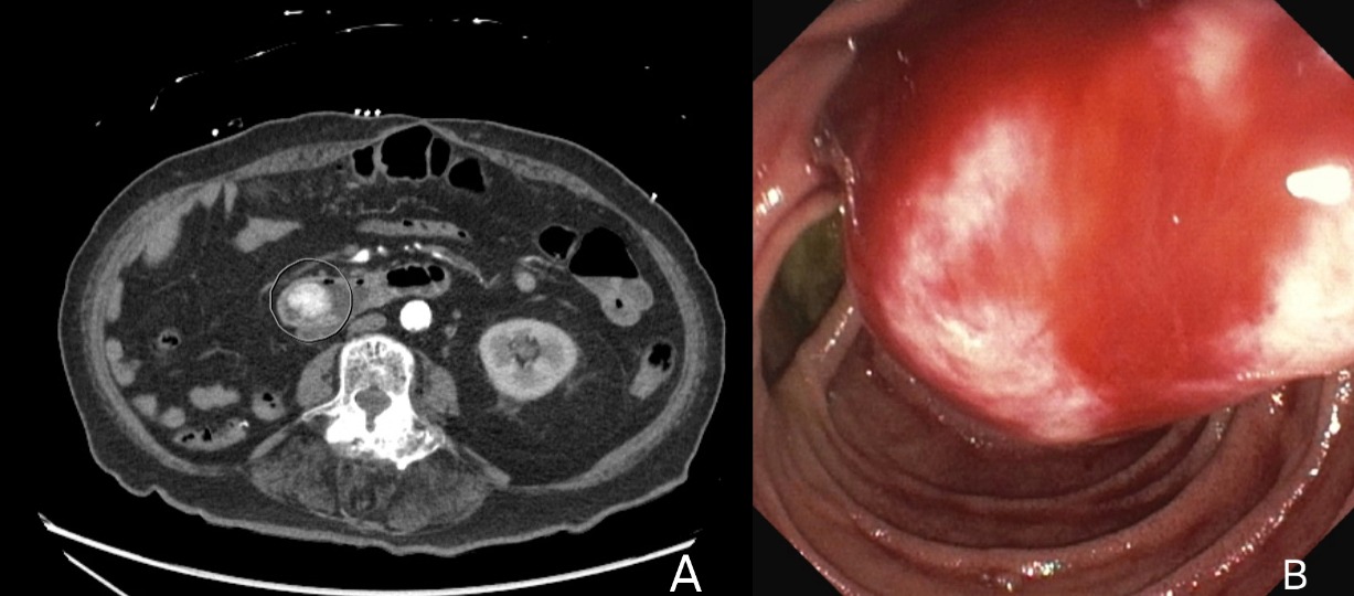
Initial hemoglobin was 6.1 and she required transfusion of packed RBCs. CTA of the abdomen and pelvis showed no extravasation to suggest active bleeding, but did show an enhancing, hyper-vascular mass in the proximal duodenum, suspicious for malignancy, image A.
Upper endoscopy showed a large, ulcerated mass with oozing on contact in the second portion of the duodenum, image B. Colonoscopy showed diverticulosis in the sigmoid colon, descending colon, and in the transverse colon.
Biopsy of the duodenal mass revealed mucosa with ulceration and acute fibrinopurulent exudate, mostly replaced by a malignant neoplasm with clear cell features. These findings confirmed a clear cell carcinoma in favor of metastasis from primary renal carcinoma to the duodenum.
Discussion: Although the precise process of RCC spreading to the duodenum remains unclear, gastrointestinal symptoms in RCC patients should prompt consideration for potential metastatic involvement. A thorough examination is warranted to explore this possibility.

Figure: Image A: CTA of the abdomen and pelvis demonstrating an enhancing, hyper-vascular mass in the proximal duodenum, suspicious for malignancy.
Image B: Upper endoscopy showed a large, ulcerated mass with oozing on contact in the second portion of the duodenum.
Image B: Upper endoscopy showed a large, ulcerated mass with oozing on contact in the second portion of the duodenum.
Disclosures:
Sana Rabeeah indicated no relevant financial relationships.
Sahithi Chinnam indicated no relevant financial relationships.
Ahmed El Rahyel indicated no relevant financial relationships.
Ali Wakil indicated no relevant financial relationships.
Hasan Al-Obaidi indicated no relevant financial relationships.
Omar Saab indicated no relevant financial relationships.
Sami Ghazaleh indicated no relevant financial relationships.
Yaseen Alastal indicated no relevant financial relationships.
Sana Rabeeah, MD1, Sahithi Chinnam, DO1, Ahmed El Rahyel, MD1, Ali Wakil, MD2, Hasan Al-Obaidi, MD3, Omar Saab, MD4, Sami Ghazaleh, MD5, Yaseen Alastal, MD6. P3309 - Rare Case of Metastatic Renal Cell Carcinoma to the Duodenum, ACG 2024 Annual Scientific Meeting Abstracts. Philadelphia, PA: American College of Gastroenterology.
