Monday Poster Session
Category: Stomach
P3386 - Unusual Presentation of Gastric Mucosa-Associated Lymphoid Tissue Lymphoma
Monday, October 28, 2024
10:30 AM - 4:00 PM ET
Location: Exhibit Hall E

Has Audio

Parvir Aujla, MD
University of Texas Health, McGovern Medical School
Houston, TX
Presenting Author(s)
Parvir Aujla, MD1, Rohit Khanna, DO2, Alex Prevallet, DO3, Abdullah S. Aleem, MD4, Faizi Hai, MD5
1University of Texas Health, McGovern Medical School, Houston, TX; 2Scripps Green Hospital, San Diego, CA; 3HCA Riverside Community Hospital, Riverside, CA; 4University of Texas at Houston, Houston, TX; 5Scripps Mercy Hospital, San Diego, CA
Introduction: Gastric mucosa-associated lymphoid tissue (MALT) lymphoma typically presents with non-specific upper gastrointestinal (GI) symptoms and common endoscopic findings. Here, we present a case of MALT lymphoma with an atypical presentation, both symptomatically and endoscopically, in a patient who presented following a ground-level fall.
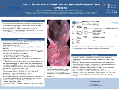
Case Description/Methods: An 80-year-old female with a history of GERD and alcohol use disorder presented after a mechanical ground-level fall and developed acute diarrhea. She reported a 6-month history of decreased appetite and a 20-pound weight loss. She was taking pantoprazole and calcium carbonate daily for acid reflux. She denied dysphagia, abdominal pain, melena, hematochezia, NSAID use, fevers, chills, night sweats, or prior EGD. Colonoscopy three years prior was normal. Labs showed a hemoglobin of 11.2 and MCV of 101. Stool studies were negative for C. difficile, enteric pathogens, WBCs, and H. pylori. CT abdomen and pelvis revealed multiple upper abdominal lymph nodes, non-specific thickening of the stomach, liquid stool throughout the GI tract, splenomegaly (15.5 cm), and prominent mediastinal lymph nodes. EGD showed atrophic mucosa within the stomach antrum and thickened, firm-appearing folds within the stomach body. There were a few 1-2 mm scattered non-bleeding erosions in the stomach, with an abundant amount of thick mucus throughout, suggestive of a protein losing enteropathy (Figure 1). The esophagus and duodenum appeared normal. Biopsies of the gastric body and antrum showed atypical lymphoid tissue consistent with MALT lymphoma, active gastritis with focal intestinal metaplasia, and rare positivity for the H. pylori stain. Duodenal biopsies were normal. Patient was ultimately treated with triple therapy and rituximab.
Discussion: Endoscopic findings in MALT lymphoma typically include small, benign-appearing ulcers, fold thickening, mucosal nodularity, and masses in high-grade disease. In our case, the gastric mucosa was atrophic and the folds contained thick mucus secretions throughout the stomach which endoscopically appeared as a protein losing enteropathy rather than a lymphoma. Patient was treated with antibiotics, for assurance of H. pylorieradication, and rituximab given the aggressive stage of her disease. Given the variable presentation of MALT lymphoma on endoscopy, our case highlights the importance of performing multiple biopsies from both normal and abnormal-appearing mucosa in patients with abnormal endoscopic findings.

Disclosures:
Parvir Aujla, MD1, Rohit Khanna, DO2, Alex Prevallet, DO3, Abdullah S. Aleem, MD4, Faizi Hai, MD5. P3386 - Unusual Presentation of Gastric Mucosa-Associated Lymphoid Tissue Lymphoma, ACG 2024 Annual Scientific Meeting Abstracts. Philadelphia, PA: American College of Gastroenterology.
1University of Texas Health, McGovern Medical School, Houston, TX; 2Scripps Green Hospital, San Diego, CA; 3HCA Riverside Community Hospital, Riverside, CA; 4University of Texas at Houston, Houston, TX; 5Scripps Mercy Hospital, San Diego, CA
Introduction: Gastric mucosa-associated lymphoid tissue (MALT) lymphoma typically presents with non-specific upper gastrointestinal (GI) symptoms and common endoscopic findings. Here, we present a case of MALT lymphoma with an atypical presentation, both symptomatically and endoscopically, in a patient who presented following a ground-level fall.
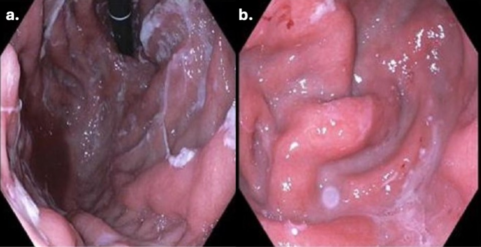
Case Description/Methods: An 80-year-old female with a history of GERD and alcohol use disorder presented after a mechanical ground-level fall and developed acute diarrhea. She reported a 6-month history of decreased appetite and a 20-pound weight loss. She was taking pantoprazole and calcium carbonate daily for acid reflux. She denied dysphagia, abdominal pain, melena, hematochezia, NSAID use, fevers, chills, night sweats, or prior EGD. Colonoscopy three years prior was normal. Labs showed a hemoglobin of 11.2 and MCV of 101. Stool studies were negative for C. difficile, enteric pathogens, WBCs, and H. pylori. CT abdomen and pelvis revealed multiple upper abdominal lymph nodes, non-specific thickening of the stomach, liquid stool throughout the GI tract, splenomegaly (15.5 cm), and prominent mediastinal lymph nodes. EGD showed atrophic mucosa within the stomach antrum and thickened, firm-appearing folds within the stomach body. There were a few 1-2 mm scattered non-bleeding erosions in the stomach, with an abundant amount of thick mucus throughout, suggestive of a protein losing enteropathy (Figure 1). The esophagus and duodenum appeared normal. Biopsies of the gastric body and antrum showed atypical lymphoid tissue consistent with MALT lymphoma, active gastritis with focal intestinal metaplasia, and rare positivity for the H. pylori stain. Duodenal biopsies were normal. Patient was ultimately treated with triple therapy and rituximab.
Discussion: Endoscopic findings in MALT lymphoma typically include small, benign-appearing ulcers, fold thickening, mucosal nodularity, and masses in high-grade disease. In our case, the gastric mucosa was atrophic and the folds contained thick mucus secretions throughout the stomach which endoscopically appeared as a protein losing enteropathy rather than a lymphoma. Patient was treated with antibiotics, for assurance of H. pylorieradication, and rituximab given the aggressive stage of her disease. Given the variable presentation of MALT lymphoma on endoscopy, our case highlights the importance of performing multiple biopsies from both normal and abnormal-appearing mucosa in patients with abnormal endoscopic findings.

Figure: Figure 1: Endoscopy showing thickened gastric folds with 1-2 mm scattered non-bleeding erosions (a.) and abundant thick mucus (b.)
Disclosures:
Parvir Aujla indicated no relevant financial relationships.
Rohit Khanna indicated no relevant financial relationships.
Alex Prevallet indicated no relevant financial relationships.
Abdullah Aleem indicated no relevant financial relationships.
Faizi Hai indicated no relevant financial relationships.
Parvir Aujla, MD1, Rohit Khanna, DO2, Alex Prevallet, DO3, Abdullah S. Aleem, MD4, Faizi Hai, MD5. P3386 - Unusual Presentation of Gastric Mucosa-Associated Lymphoid Tissue Lymphoma, ACG 2024 Annual Scientific Meeting Abstracts. Philadelphia, PA: American College of Gastroenterology.
