Monday Poster Session
Category: Stomach
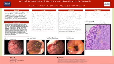
P3407 - An Unfortunate Case of Breast Cancer Metastasis to the Stomach
Monday, October 28, 2024
10:30 AM - 4:00 PM ET
Location: Exhibit Hall E

Has Audio

Tatsiana Serhiyenia, MD
Einstein Healthcare Network
Bryn Mawr, PA
Presenting Author(s)
Tatsiana Serhiyenia, MD1, Matthew Chan, DO2, Steven Stanek, MD2, Mark Morginstin, DO3, Julianne N. Qualtieri, MD2
1Einstein Healthcare Network, Bryn Mawr, PA; 2Einstein Healthcare Network, East Norriton, PA; 3Einstein Healthcare Network, Ambler, PA
Introduction: Breast cancer is the most common cancer among females worldwide and the second-leading cause of cancer death.
Despite increased awareness and screening practice, it often discovered at later stages. Even when diagnosed early, it can metastasize a few years after. The bones, lungs, brain, liver are the most common metastases sites. Only 0.3% in retrospective studies and 8–18% in autopsy were found in the GI tract, most commonly from invasive lobular carcinoma. We present a case of gastric metastases of lobular carcinoma 4 years after breast cancer diagnosis.
Case Description/Methods: A 59-year-old female with right lobular carcinoma diagnosed in 2020, post bilateral mastectomy, chemotherapy, radiation, on Anastrozole, presented with abdominal distention, dyspnea on exertion, pedal edema for three weeks.
Denied nausea, vomiting, GI bleeding. Vitals were notable for normotension, tachycardia, mild tachypnea. Exam revealed distended abdomen, epigastric pain on palpation. Labs showed elevated creatinine, mild transaminitis.
CT abdomen showed large ascites, omental nodular thickening, gastric wall enhancement, suspected bony metastases. Thora- and paracentesis were done. Cytology revealed metastatic mammary carcinoma ER positive and HER2 negative.
Gastroenterology consulted. EGD revealed diffuse, severe mucosal erythema, erosion, friability with spontaneous bleeding, granularity, inflammation, moderate stenosis in the prepyloric region of the stomach.
Abnormal gastric mucosa biopsy described extensive infiltration of lamina propria by metastatic lobular breast carcinoma.
Discussion: Gastric metastases from breast cancer are rare. Invasive lobular carcinoma is most common culprit.
Symptoms vary from non-specific (nausea, decreased appetite, bloating), to specific (vomiting, weight loss, bleeding). Asymptomatic cases exist. Specific symptoms raise suspicion of primary stomach cancer, necessitating prompt endoscopic biopsy for diagnosis.
Metastatic breast cancer has diverse endoscopic appearances: discolored mucosal lesions, polyps with or without ulcerations, diffuse infiltrative lesion with bleeding, fold thickening and deep ulceration with bleeding. It can appear in any stomach region. Submucosal and seromuscular make 50% of gastric metastases and may not show on endoscopy, requiring deep tissue biopsy for diagnosis.
Our case motivates all clinicians to suspect GI metastasis in breast cancer patients with GI symptoms. Timely endoscopic evaluation and proper diagnosis aid in proper treatment.

Disclosures:
Tatsiana Serhiyenia, MD1, Matthew Chan, DO2, Steven Stanek, MD2, Mark Morginstin, DO3, Julianne N. Qualtieri, MD2. P3407 - An Unfortunate Case of Breast Cancer Metastasis to the Stomach, ACG 2024 Annual Scientific Meeting Abstracts. Philadelphia, PA: American College of Gastroenterology.
1Einstein Healthcare Network, Bryn Mawr, PA; 2Einstein Healthcare Network, East Norriton, PA; 3Einstein Healthcare Network, Ambler, PA
Introduction: Breast cancer is the most common cancer among females worldwide and the second-leading cause of cancer death.
Despite increased awareness and screening practice, it often discovered at later stages. Even when diagnosed early, it can metastasize a few years after. The bones, lungs, brain, liver are the most common metastases sites. Only 0.3% in retrospective studies and 8–18% in autopsy were found in the GI tract, most commonly from invasive lobular carcinoma. We present a case of gastric metastases of lobular carcinoma 4 years after breast cancer diagnosis.
Case Description/Methods: A 59-year-old female with right lobular carcinoma diagnosed in 2020, post bilateral mastectomy, chemotherapy, radiation, on Anastrozole, presented with abdominal distention, dyspnea on exertion, pedal edema for three weeks.
Denied nausea, vomiting, GI bleeding. Vitals were notable for normotension, tachycardia, mild tachypnea. Exam revealed distended abdomen, epigastric pain on palpation. Labs showed elevated creatinine, mild transaminitis.
CT abdomen showed large ascites, omental nodular thickening, gastric wall enhancement, suspected bony metastases. Thora- and paracentesis were done. Cytology revealed metastatic mammary carcinoma ER positive and HER2 negative.
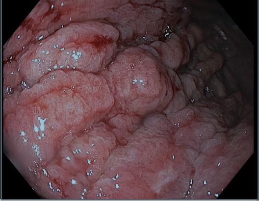
Gastroenterology consulted. EGD revealed diffuse, severe mucosal erythema, erosion, friability with spontaneous bleeding, granularity, inflammation, moderate stenosis in the prepyloric region of the stomach.
Abnormal gastric mucosa biopsy described extensive infiltration of lamina propria by metastatic lobular breast carcinoma.
Discussion: Gastric metastases from breast cancer are rare. Invasive lobular carcinoma is most common culprit.
Symptoms vary from non-specific (nausea, decreased appetite, bloating), to specific (vomiting, weight loss, bleeding). Asymptomatic cases exist. Specific symptoms raise suspicion of primary stomach cancer, necessitating prompt endoscopic biopsy for diagnosis.
Metastatic breast cancer has diverse endoscopic appearances: discolored mucosal lesions, polyps with or without ulcerations, diffuse infiltrative lesion with bleeding, fold thickening and deep ulceration with bleeding. It can appear in any stomach region. Submucosal and seromuscular make 50% of gastric metastases and may not show on endoscopy, requiring deep tissue biopsy for diagnosis.
Our case motivates all clinicians to suspect GI metastasis in breast cancer patients with GI symptoms. Timely endoscopic evaluation and proper diagnosis aid in proper treatment.

Figure: Gastric Body
Disclosures:
Tatsiana Serhiyenia indicated no relevant financial relationships.
Matthew Chan indicated no relevant financial relationships.
Steven Stanek indicated no relevant financial relationships.
Mark Morginstin indicated no relevant financial relationships.
Julianne Qualtieri indicated no relevant financial relationships.
Tatsiana Serhiyenia, MD1, Matthew Chan, DO2, Steven Stanek, MD2, Mark Morginstin, DO3, Julianne N. Qualtieri, MD2. P3407 - An Unfortunate Case of Breast Cancer Metastasis to the Stomach, ACG 2024 Annual Scientific Meeting Abstracts. Philadelphia, PA: American College of Gastroenterology.
