Monday Poster Session
Category: Stomach
P3413 - A Rare Case of Metastatic Hepatoid Adenocarcinoma of the Stomach
Monday, October 28, 2024
10:30 AM - 4:00 PM ET
Location: Exhibit Hall E

Has Audio
- PY
Patrick Yang, MD
Westchester Medical Center
Valhalla, NY
Presenting Author(s)
Patrick Yang, MD, Liying Han, MD, Agnieszka Nagorna, MD, Cynthia Cohen, MD
Westchester Medical Center, Valhalla, NY
Introduction: Hepatoid adenocarcinoma of the stomach (HAS) is a rare, aggressive, malignant tumor, accounting for 0.17%-15% of gastric cancers. It is morphologically identical to hepatocellular carcinoma (HCC), however presents in the stomach instead of the liver. The clinical manifestations are often nonspecific and similar to other gastric cancers. Here, we report a rare case of gastric hepatoid adenocarcinoma in a patient presented with abdominal pain and GI bleed.
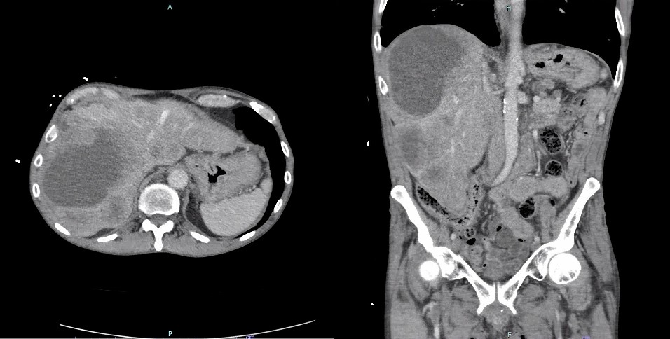
Case Description/Methods: 59-year-old male smoker with past medical history of COPD, diverticulitis requiring a colostomy with revision, and alcohol use disorder presented with abdominal pain. CT abdomen revealed multiple hepatic lesions, the being a right-sided largest measuring 12.7 x 8.6 cm with concerns for necrosis and metastatic disease. There also was an irregular appearance of the stomach, concerning for an underlying mass with surrounding necrotic lymph nodes. Serum AFP was elevated to over 20,000 ng/ml. The patient had two episode of coffee-ground emesis and IV pantoprazole was initiated. EGD on the following day revealed an ulcerated mass in the gastric cardia, and biopsies were taken. Biopsy revealed a moderately differentiated malignant neoplasm of the gastric mucosa with immunohistochemical staining positive for AFP, Glypican 3, SALL4, PLAP, CK19, CK18, CDX2, MOC31, BerEp4, and focal positivity for Hepa1. It was negative for PDL-1 and Her2. The differential diagnosis included gastric hepatoid adenocarcinoma with metastasis to the liver, versus primary yolk sac tumor of the liver metastatic to the stomach, versus gastric yok sac tumor with liver metastasis. Outside consultation to the NIH pathology lab was consistent with AFP-producing hepatoid gastric adenocarcinoma. MRI brain did not show any metastases and the patient was discharged with oncology follow-up.
Discussion: Gastric hepatoid adenocarcinomas are aggressive and often diagnosed at an advanced stage when they have already metastasized to the liver, lymph nodes, or other tissues. They typically have a worse prognosis than other gastric cancers and there is no consensus regarding therapy. As they are similar in morphology to HCC, they generally produce high levels of AFP. When gastric hepatoid adenocarcinomas metastasize to the liver, they may be misdiagnosed as HCC, especially if only a single or few hepatic lesions are discovered. Physicians should be vigilant with patients found to have multiple hepatic lesions with high AFP and no history of hepatitis or cirrhosis.

Disclosures:
Patrick Yang, MD, Liying Han, MD, Agnieszka Nagorna, MD, Cynthia Cohen, MD. P3413 - A Rare Case of Metastatic Hepatoid Adenocarcinoma of the Stomach, ACG 2024 Annual Scientific Meeting Abstracts. Philadelphia, PA: American College of Gastroenterology.
Westchester Medical Center, Valhalla, NY
Introduction: Hepatoid adenocarcinoma of the stomach (HAS) is a rare, aggressive, malignant tumor, accounting for 0.17%-15% of gastric cancers. It is morphologically identical to hepatocellular carcinoma (HCC), however presents in the stomach instead of the liver. The clinical manifestations are often nonspecific and similar to other gastric cancers. Here, we report a rare case of gastric hepatoid adenocarcinoma in a patient presented with abdominal pain and GI bleed.
Case Description/Methods: 59-year-old male smoker with past medical history of COPD, diverticulitis requiring a colostomy with revision, and alcohol use disorder presented with abdominal pain. CT abdomen revealed multiple hepatic lesions, the being a right-sided largest measuring 12.7 x 8.6 cm with concerns for necrosis and metastatic disease. There also was an irregular appearance of the stomach, concerning for an underlying mass with surrounding necrotic lymph nodes. Serum AFP was elevated to over 20,000 ng/ml. The patient had two episode of coffee-ground emesis and IV pantoprazole was initiated. EGD on the following day revealed an ulcerated mass in the gastric cardia, and biopsies were taken. Biopsy revealed a moderately differentiated malignant neoplasm of the gastric mucosa with immunohistochemical staining positive for AFP, Glypican 3, SALL4, PLAP, CK19, CK18, CDX2, MOC31, BerEp4, and focal positivity for Hepa1. It was negative for PDL-1 and Her2. The differential diagnosis included gastric hepatoid adenocarcinoma with metastasis to the liver, versus primary yolk sac tumor of the liver metastatic to the stomach, versus gastric yok sac tumor with liver metastasis. Outside consultation to the NIH pathology lab was consistent with AFP-producing hepatoid gastric adenocarcinoma. MRI brain did not show any metastases and the patient was discharged with oncology follow-up.
Discussion: Gastric hepatoid adenocarcinomas are aggressive and often diagnosed at an advanced stage when they have already metastasized to the liver, lymph nodes, or other tissues. They typically have a worse prognosis than other gastric cancers and there is no consensus regarding therapy. As they are similar in morphology to HCC, they generally produce high levels of AFP. When gastric hepatoid adenocarcinomas metastasize to the liver, they may be misdiagnosed as HCC, especially if only a single or few hepatic lesions are discovered. Physicians should be vigilant with patients found to have multiple hepatic lesions with high AFP and no history of hepatitis or cirrhosis.

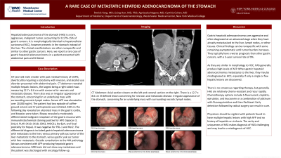
Figure: CT Abdomen: Axial section shown on the left and coronal section on the right. There is a 12.7 x 8.6 cm ill-defined lesion concerning for necrosis and metastatic disease. Irregular appearance of the stomach, concerning for an underlying mass with surrounding necrotic lymph nodes.
Disclosures:
Patrick Yang indicated no relevant financial relationships.
Liying Han indicated no relevant financial relationships.
Agnieszka Nagorna indicated no relevant financial relationships.
Cynthia Cohen indicated no relevant financial relationships.
Patrick Yang, MD, Liying Han, MD, Agnieszka Nagorna, MD, Cynthia Cohen, MD. P3413 - A Rare Case of Metastatic Hepatoid Adenocarcinoma of the Stomach, ACG 2024 Annual Scientific Meeting Abstracts. Philadelphia, PA: American College of Gastroenterology.
