Monday Poster Session
Category: Stomach
P3415 - Venturing Into the Varied Realm of Varioliform Gastritis: Unveiling an Unconventional Culprit Behind Rare Gastric Lesions
Monday, October 28, 2024
10:30 AM - 4:00 PM ET
Location: Exhibit Hall E

Has Audio

Tevan Luong, BS
University of California, Irvine
Orange, CA
Presenting Author(s)
Hien Lau, MD, Rishi Vermani, BS, Tevan Luong, BS, C Gregory Albers, MD
University of California, Irvine, Orange, CA
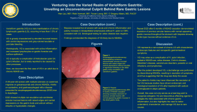
Introduction: Varioliform gastritis (VG) is a rare manifestation of chronic lymphocytic gastritis (LG), occurring in less than 1.5% of cases. VG is grossly characterized by elevated mucosal lesions with central depressions and gray-colored exudate or punctate bleeding. Histologically, VG is associated with active inflammation and increased lymphocytes on gastric foveolar and surface epithelium. VG is typically a complication of Helicobacter pylori (H. pylori) infection, but is rarely reported to be caused by other processes. Here, we document the first case of VG in an adult due to chronic NSAID use.
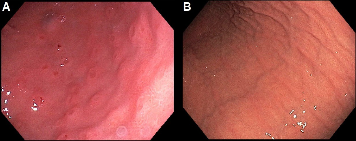
Case Description/Methods: A 49-year-old woman with multiple sclerosis on ozanimod, migraine on galcanezumab and chronic NSAIDs, rhinitis on azelastine, and gastroesophageal reflux disease presented for esophagogastroduodenoscopy (EGD) due to dysphagia. EGD showed multiple non-exudative subcentimeter annular mucosal lesions with raised edges and central depressions on the gastric body and antrum without atrophic or hyperplastic changes. Biopsies revealed foci of erosion with acute and chronic inflammation and patchy increase in intraepithelial lymphocytes without H. pylori or CMV, consistent with LG. Serological testing for celiac disease was also negative. Findings corroborated the diagnosis of VG likely due to NSAIDs. Repeat EGD after 6 months of NSAID cessation demonstrated resolution of previous annular lesions with normal appearing gastric mucosa throughout the stomach with biopsies showing fundic-type mucosa without evidence of LG.
Discussion: VG represents a rare subset of chronic LG with characteristic endoscopic features and nonspecific gastrointestinal symptoms. While VG may be a complication of H. pylori infection, rare cases associated with NSAID use in pediatric patients, celiac disease, Crohn’s disease, Ménétrier’s disease, autoimmune disorders, parasitic or viral infections, and lymphomas have been reported. A relationship between VG and atopy, such as asthma and allergic rhinitis, has been suggested with studies showing endoscopic and histological resolution of VG after treatment with sodium cromoglycate in atopic patients. Based on these trials, the patient’s rhinitis may have played a role in the course of their VG. Overall, this case not only serves as a learning point to recognize iatrogenic VG and discontinue offending agents to mitigate the risks associated with chronic gastric inflammation, but also highlights the need to better understand, characterize, and manage VG due to rare causes.

Disclosures:
Hien Lau, MD, Rishi Vermani, BS, Tevan Luong, BS, C Gregory Albers, MD. P3415 - Venturing Into the Varied Realm of Varioliform Gastritis: Unveiling an Unconventional Culprit Behind Rare Gastric Lesions, ACG 2024 Annual Scientific Meeting Abstracts. Philadelphia, PA: American College of Gastroenterology.
University of California, Irvine, Orange, CA
Introduction: Varioliform gastritis (VG) is a rare manifestation of chronic lymphocytic gastritis (LG), occurring in less than 1.5% of cases. VG is grossly characterized by elevated mucosal lesions with central depressions and gray-colored exudate or punctate bleeding. Histologically, VG is associated with active inflammation and increased lymphocytes on gastric foveolar and surface epithelium. VG is typically a complication of Helicobacter pylori (H. pylori) infection, but is rarely reported to be caused by other processes. Here, we document the first case of VG in an adult due to chronic NSAID use.
Case Description/Methods: A 49-year-old woman with multiple sclerosis on ozanimod, migraine on galcanezumab and chronic NSAIDs, rhinitis on azelastine, and gastroesophageal reflux disease presented for esophagogastroduodenoscopy (EGD) due to dysphagia. EGD showed multiple non-exudative subcentimeter annular mucosal lesions with raised edges and central depressions on the gastric body and antrum without atrophic or hyperplastic changes. Biopsies revealed foci of erosion with acute and chronic inflammation and patchy increase in intraepithelial lymphocytes without H. pylori or CMV, consistent with LG. Serological testing for celiac disease was also negative. Findings corroborated the diagnosis of VG likely due to NSAIDs. Repeat EGD after 6 months of NSAID cessation demonstrated resolution of previous annular lesions with normal appearing gastric mucosa throughout the stomach with biopsies showing fundic-type mucosa without evidence of LG.
Discussion: VG represents a rare subset of chronic LG with characteristic endoscopic features and nonspecific gastrointestinal symptoms. While VG may be a complication of H. pylori infection, rare cases associated with NSAID use in pediatric patients, celiac disease, Crohn’s disease, Ménétrier’s disease, autoimmune disorders, parasitic or viral infections, and lymphomas have been reported. A relationship between VG and atopy, such as asthma and allergic rhinitis, has been suggested with studies showing endoscopic and histological resolution of VG after treatment with sodium cromoglycate in atopic patients. Based on these trials, the patient’s rhinitis may have played a role in the course of their VG. Overall, this case not only serves as a learning point to recognize iatrogenic VG and discontinue offending agents to mitigate the risks associated with chronic gastric inflammation, but also highlights the need to better understand, characterize, and manage VG due to rare causes.

Figure: Figure: A) Esophagogastroduodenoscopy demonstrating numerous subcentimeter annular mucosal lesions with raised edges and central depressions without exudate or significant bleeding in the distal gastric body consistent with varioliform gastritis. B) Repeat esophagogastroduodenoscopy after 6 months of NSAID cessation showing complete resolution of previous annular lesions with normal appearing gastric mucosa in the distal gastric body.
Disclosures:
Hien Lau indicated no relevant financial relationships.
Rishi Vermani indicated no relevant financial relationships.
Tevan Luong indicated no relevant financial relationships.
C Gregory Albers: Nestle Health – Speakers Bureau. Phathom Pharmaceuticals – Speakers Bureau. Salix Pharmaceuticals – Speakers Bureau.
Hien Lau, MD, Rishi Vermani, BS, Tevan Luong, BS, C Gregory Albers, MD. P3415 - Venturing Into the Varied Realm of Varioliform Gastritis: Unveiling an Unconventional Culprit Behind Rare Gastric Lesions, ACG 2024 Annual Scientific Meeting Abstracts. Philadelphia, PA: American College of Gastroenterology.
