Tuesday Poster Session
Category: Biliary/Pancreas
P3448 - Endoscopic Ultrasound Features of Pancreatic Tuberculosis - A Case Series From Tuberculosis Endemic Country
Tuesday, October 29, 2024
10:30 AM - 4:00 PM ET
Location: Exhibit Hall E

Has Audio

Aditya P. Kale, DM
Advance Center for Treatment Research and Education in Cancer- Tata Memorial Center
Navi Mumbai, Maharashtra, India
Presenting Author(s)
Aditya P. Kale, DM1, Unique Tyagi, DM2, Shravan GH, MD2, Akhil Mahajan, MD2, Rahul Puri, MD2, Aruna ravi, MD2, Sandip supnar, MD2, Kiran Mane, DM2, Aadish Kumar Jain, DM2, Sridhar Sundaram, DM2, Prachi S. Patil, DM2, Shaesta A. Mehta, DM3
1Advance Center for Treatment Research and Education in Cancer- Tata Memorial Center, Navi Mumbai, Maharashtra, India; 2Tata Memorial Hospital, Mumbai, Maharashtra, India; 3Tata Memorial Hospitald, Mumbai, Maharashtra, India
Introduction: Diagnosis of pancreatic tuberculosis (PTB) is challenging due to lack of specific clinical & imaging features.
Methods: We retrospectively studied 20 cases of PTB, evaluated at our center between 2019-2024. Demography, clinical details, laboratory investigations, radiology, endoscopic ultrasound (EUS) and histology findings were noted.
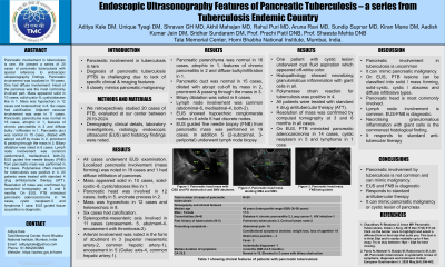
Results: Table 1 shows demography, clinical and radiological findings. All cases underwent EUS. Localized pancreatic involvement (mass forming) was noted in 19 cases and 1 had diffuse infiltration of pancreas. Mass appeared solid in 10 cases, solid-cystic–8, cystic/abscess like in 1. Pancreatic head was involved in 12 cases, body in 5, uncinate process in 2. Mass was hypoechoic in 12 cases and heteroechoic in 8. Six cases had calcification. Splenoportal-mesenteric axis was involved in 11 cases (encasement- 5, abutment-4, encasement with thrombosis-2). Arterial involvement was noted in the form of abutment in 3 (superior mesenteric artery-2, common hepatic artery-1), encasement in 5 (Celiac axis-4, common hepatic artery-1). Pancreatic parenchyma was normal in 16 cases, atrophic in 1, features of chronic pancreatitis in 2 and diffuse bulky/infiltration in 1. Pancreatic duct was normal in 15 cases, dilated with abrupt cut-off by mass in 2, prominent & passing through the mass in 3. Biliary dilatation was noted in 6 cases. Lymph node involvement was common (abdominal-6, mediastinal-4, both-2). EUS showed hypoechoic conglomerate nodes in 6 while 6 had discrete nodes. EUS guided fine needle biopsy (FNB) from pancreatic mass was performed in 19 cases. In addition 5 (2-subcarinal, 3-periportal) underwent lymph node biopsy. One patient with cystic lesion underwent cyst fluid aspiration which appeared off-white colour. Histopathology showed necrotising granulomatous inflammation with giant cells in all. Polymerase chain reaction for tuberculosis was positive in 4. All patients were treated with standard 4 drug antitubercular therapy (ATT). Resolution of mass was confirmed by computed tomography at 3 and 6 months. On EUS, PTB mimicked pancreatic adenocarcinoma in 14 cases, cystic neoplasm-5 and lymphoma 1 case.
Discussion: PTB mimics pancreatic malignancy. On EUS, PTB lesions can be classified into solid/mass forming, solid-cystic, cystic/abscess and diffuse infiltrative types. Pancreatic head is most commonly involved. Lymph node involvement is common. EUS-FNB is diagnostic. It responds to standard ATT.
Note: The table for this abstract can be viewed in the ePoster Gallery section of the ACG 2024 ePoster Site or in The American Journal of Gastroenterology's abstract supplement issue, both of which will be available starting October 27, 2024.
Disclosures:
Aditya P. Kale, DM1, Unique Tyagi, DM2, Shravan GH, MD2, Akhil Mahajan, MD2, Rahul Puri, MD2, Aruna ravi, MD2, Sandip supnar, MD2, Kiran Mane, DM2, Aadish Kumar Jain, DM2, Sridhar Sundaram, DM2, Prachi S. Patil, DM2, Shaesta A. Mehta, DM3. P3448 - Endoscopic Ultrasound Features of Pancreatic Tuberculosis - A Case Series From Tuberculosis Endemic Country, ACG 2024 Annual Scientific Meeting Abstracts. Philadelphia, PA: American College of Gastroenterology.
1Advance Center for Treatment Research and Education in Cancer- Tata Memorial Center, Navi Mumbai, Maharashtra, India; 2Tata Memorial Hospital, Mumbai, Maharashtra, India; 3Tata Memorial Hospitald, Mumbai, Maharashtra, India
Introduction: Diagnosis of pancreatic tuberculosis (PTB) is challenging due to lack of specific clinical & imaging features.
Methods: We retrospectively studied 20 cases of PTB, evaluated at our center between 2019-2024. Demography, clinical details, laboratory investigations, radiology, endoscopic ultrasound (EUS) and histology findings were noted.
Results: Table 1 shows demography, clinical and radiological findings. All cases underwent EUS. Localized pancreatic involvement (mass forming) was noted in 19 cases and 1 had diffuse infiltration of pancreas. Mass appeared solid in 10 cases, solid-cystic–8, cystic/abscess like in 1. Pancreatic head was involved in 12 cases, body in 5, uncinate process in 2. Mass was hypoechoic in 12 cases and heteroechoic in 8. Six cases had calcification. Splenoportal-mesenteric axis was involved in 11 cases (encasement- 5, abutment-4, encasement with thrombosis-2). Arterial involvement was noted in the form of abutment in 3 (superior mesenteric artery-2, common hepatic artery-1), encasement in 5 (Celiac axis-4, common hepatic artery-1). Pancreatic parenchyma was normal in 16 cases, atrophic in 1, features of chronic pancreatitis in 2 and diffuse bulky/infiltration in 1. Pancreatic duct was normal in 15 cases, dilated with abrupt cut-off by mass in 2, prominent & passing through the mass in 3. Biliary dilatation was noted in 6 cases. Lymph node involvement was common (abdominal-6, mediastinal-4, both-2). EUS showed hypoechoic conglomerate nodes in 6 while 6 had discrete nodes. EUS guided fine needle biopsy (FNB) from pancreatic mass was performed in 19 cases. In addition 5 (2-subcarinal, 3-periportal) underwent lymph node biopsy. One patient with cystic lesion underwent cyst fluid aspiration which appeared off-white colour. Histopathology showed necrotising granulomatous inflammation with giant cells in all. Polymerase chain reaction for tuberculosis was positive in 4. All patients were treated with standard 4 drug antitubercular therapy (ATT). Resolution of mass was confirmed by computed tomography at 3 and 6 months. On EUS, PTB mimicked pancreatic adenocarcinoma in 14 cases, cystic neoplasm-5 and lymphoma 1 case.
Discussion: PTB mimics pancreatic malignancy. On EUS, PTB lesions can be classified into solid/mass forming, solid-cystic, cystic/abscess and diffuse infiltrative types. Pancreatic head is most commonly involved. Lymph node involvement is common. EUS-FNB is diagnostic. It responds to standard ATT.
Note: The table for this abstract can be viewed in the ePoster Gallery section of the ACG 2024 ePoster Site or in The American Journal of Gastroenterology's abstract supplement issue, both of which will be available starting October 27, 2024.
Disclosures:
Aditya Kale indicated no relevant financial relationships.
Unique Tyagi indicated no relevant financial relationships.
Shravan GH indicated no relevant financial relationships.
Akhil Mahajan indicated no relevant financial relationships.
Rahul Puri indicated no relevant financial relationships.
Aruna ravi indicated no relevant financial relationships.
Sandip supnar indicated no relevant financial relationships.
Kiran Mane indicated no relevant financial relationships.
Aadish Kumar Jain indicated no relevant financial relationships.
Sridhar Sundaram indicated no relevant financial relationships.
Prachi Patil indicated no relevant financial relationships.
Shaesta Mehta indicated no relevant financial relationships.
Aditya P. Kale, DM1, Unique Tyagi, DM2, Shravan GH, MD2, Akhil Mahajan, MD2, Rahul Puri, MD2, Aruna ravi, MD2, Sandip supnar, MD2, Kiran Mane, DM2, Aadish Kumar Jain, DM2, Sridhar Sundaram, DM2, Prachi S. Patil, DM2, Shaesta A. Mehta, DM3. P3448 - Endoscopic Ultrasound Features of Pancreatic Tuberculosis - A Case Series From Tuberculosis Endemic Country, ACG 2024 Annual Scientific Meeting Abstracts. Philadelphia, PA: American College of Gastroenterology.
