Tuesday Poster Session
Category: Biliary/Pancreas
P3535 - Acute Myeloid Leukemia Presenting With Leukemic Infiltration of the Liver and Bile Ducts
Tuesday, October 29, 2024
10:30 AM - 4:00 PM ET
Location: Exhibit Hall E

Has Audio

Melanie Cabezas, MD
University of South Florida
Tampa, FL
Presenting Author(s)
Award: Presidential Poster Award
Melanie Cabezas, MD, Jonathan Hilal, MD
University of South Florida, Tampa, FL
Introduction: Acute myeloid leukemia (AML) is a common hematologic malignancy characterized by the proliferation of myeloid blasts that fail to undergo normal differentiation. Although clinical presentation can include extramedullary findings of gastrointestinal infiltration, biliary obstruction is rare.
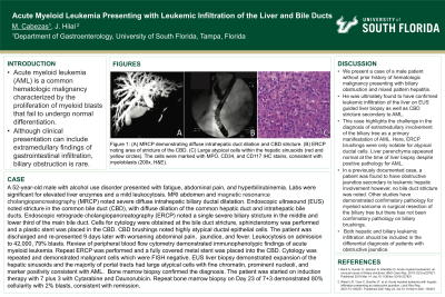
Case Description/Methods: A 52-year-old male with alcohol use disorder presented to the hospital with fatigue, epigastric abdominal pain, and hyperbilirubinemia. Labs were significant for elevated liver enzymes and a mild leukocytosis. MRI abdomen and magnetic resonance cholangiopancreatography (MRCP) were performed and noted severe diffuse intrahepatic biliary ductal dilatation. Endoscopic ultrasound (EUS) noted stricture in the common bile duct (CBD), dilation in the common hepatic duct and dilation in the intrahepatic bile ducts, diffusely. Endoscopic retrograde cholangiopancreatography (ERCP) noted a single severe biliary stricture in the middle and lower third of the main bile duct. Cells for cytology were obtained at the bile duct stricture, sphincterotomy was performed and a plastic stent was placed in CBD. CBD brushings noted highly atypical ductal epithelial cells. The patient was discharged home.
The patient presented 9 days after discharge with worsening abdominal pain, jaundice, and fever. Labs on admission notable for leukocytosis to 42,000, 79% blasts. Review of peripheral blood flow cytometry demonstrated immunophenotypic findings of acute myeloid leukemia. Repeat ERCP was performed and a fully covered metal stent was placed into the CBD. Cytology was repeated and demonstrated malignant cells which were FISH negative. EUS with liver biopsy of the left and right hepatic lobes demonstrated expansion of the hepatic sinusoids and the majority of portal tracts had large atypical cells with fine chromatin, prominent nucleoli, and marker positivity consistent with AML. Bone marrow biopsy confirmed the diagnosis. The patient was started on induction therapy with 7 plus 3 with Cytarabine and Daunorubicin. Repeat bone marrow biopsy on Day 23 of 7+3 demonstrated 80% cellularity with 2% blasts, consistent with remission.
Discussion: Extramedullary involvement by acute myeloid leukemia is rare, with manifestations ranging from cellular infiltration without effacement of tissue architecture to the growth of a tumor mass, also known as myeloid sarcoma. Literature documenting either leukemic infiltration of the liver alone or that of the extrahepatic biliary tree is limited.

Disclosures:
Melanie Cabezas, MD, Jonathan Hilal, MD. P3535 - Acute Myeloid Leukemia Presenting With Leukemic Infiltration of the Liver and Bile Ducts, ACG 2024 Annual Scientific Meeting Abstracts. Philadelphia, PA: American College of Gastroenterology.
Melanie Cabezas, MD, Jonathan Hilal, MD
University of South Florida, Tampa, FL
Introduction: Acute myeloid leukemia (AML) is a common hematologic malignancy characterized by the proliferation of myeloid blasts that fail to undergo normal differentiation. Although clinical presentation can include extramedullary findings of gastrointestinal infiltration, biliary obstruction is rare.
Case Description/Methods: A 52-year-old male with alcohol use disorder presented to the hospital with fatigue, epigastric abdominal pain, and hyperbilirubinemia. Labs were significant for elevated liver enzymes and a mild leukocytosis. MRI abdomen and magnetic resonance cholangiopancreatography (MRCP) were performed and noted severe diffuse intrahepatic biliary ductal dilatation. Endoscopic ultrasound (EUS) noted stricture in the common bile duct (CBD), dilation in the common hepatic duct and dilation in the intrahepatic bile ducts, diffusely. Endoscopic retrograde cholangiopancreatography (ERCP) noted a single severe biliary stricture in the middle and lower third of the main bile duct. Cells for cytology were obtained at the bile duct stricture, sphincterotomy was performed and a plastic stent was placed in CBD. CBD brushings noted highly atypical ductal epithelial cells. The patient was discharged home.
The patient presented 9 days after discharge with worsening abdominal pain, jaundice, and fever. Labs on admission notable for leukocytosis to 42,000, 79% blasts. Review of peripheral blood flow cytometry demonstrated immunophenotypic findings of acute myeloid leukemia. Repeat ERCP was performed and a fully covered metal stent was placed into the CBD. Cytology was repeated and demonstrated malignant cells which were FISH negative. EUS with liver biopsy of the left and right hepatic lobes demonstrated expansion of the hepatic sinusoids and the majority of portal tracts had large atypical cells with fine chromatin, prominent nucleoli, and marker positivity consistent with AML. Bone marrow biopsy confirmed the diagnosis. The patient was started on induction therapy with 7 plus 3 with Cytarabine and Daunorubicin. Repeat bone marrow biopsy on Day 23 of 7+3 demonstrated 80% cellularity with 2% blasts, consistent with remission.
Discussion: Extramedullary involvement by acute myeloid leukemia is rare, with manifestations ranging from cellular infiltration without effacement of tissue architecture to the growth of a tumor mass, also known as myeloid sarcoma. Literature documenting either leukemic infiltration of the liver alone or that of the extrahepatic biliary tree is limited.

Figure: (A) MRCP demonstrating diffuse intrahepatic duct dilation and CBD stricture (B) ERCP demonstrating CBD stricture (C) High power image demonstrating large atypical cells (red and yellow circles) within the hepatic sinusoids. The cells were decorated with MPO, CD34, and CD117 IHC stains, consistent with myeloblasts (200x, H&E).
Disclosures:
Melanie Cabezas indicated no relevant financial relationships.
Jonathan Hilal indicated no relevant financial relationships.
Melanie Cabezas, MD, Jonathan Hilal, MD. P3535 - Acute Myeloid Leukemia Presenting With Leukemic Infiltration of the Liver and Bile Ducts, ACG 2024 Annual Scientific Meeting Abstracts. Philadelphia, PA: American College of Gastroenterology.

