Tuesday Poster Session
Category: Biliary/Pancreas
P3544 - The Gargantuan Gallbladder: A Case of Gastric Outlet Obstruction Caused by a Massively Dilated Stoneless Gallbladder
Tuesday, October 29, 2024
10:30 AM - 4:00 PM ET
Location: Exhibit Hall E

Has Audio

Syed Hamaad Rahman, DO
Methodist Dallas Medical Center
Irving, TX
Presenting Author(s)
Syed Hamaad Rahman, DO1, Kyle Schneider, MD2, Blake Thompson, MD3
1Methodist Dallas Medical Center, Irving, TX; 2Methodist Dallas Medical Center, Dallas, TX; 3Methodist Health System, Dallas, TX
Introduction: Gastric outlet obstruction (GOO) is usually caused by blockage from underlying cancers, especially peripancreatic neoplasms. Symptoms include nausea, vomiting, epigastric pain, early satiety, abdominal distension, and weight loss. Physical exam may reveal a palpable mass or succussion splash, though these are not highly sensative. Gallbladder (GB) mucocele is a very rare cause of GOO, with limited reports in the literature. We report a rare case of GOO caused by a massively dilated stoneless GB mucocele.
Case Description/Methods: A 62 y/o male with PMH of HTN, CAD, PVD, and recently discovered hepatic mass of unknown origin presented to the ED with progressive abdominal pain, nausea, and bilious emesis. A few months prior, an 8.2 cm necrotic left hepatic lobe mass was biopsied revealing adenocarcinoma of pancreaticobiliary origin vs less likely GI origin. PET/CT revealed a large FDG avid mass and multiple small avid lesions concerning for metastases. Hypermetabolic wall thickening at the ileocecal junction and a lymph node near GB were suggestive of colonic malignancy.
Colonoscopy was attempted but was aborted due to poor preparation secondary to severe nausea and vomiting. The symptoms worsened with dark brown non-bloody emesis, constipation, and weight loss. On arrival he was hemodynamically stable, with exam revealing cachexia, soft and tender abdomen over the mid epigastric and right upper quadrant regions, with mild distension. Initial labs were normal, but liver function tests and bilirubin up-trended throughout the hospital course.
CT Abdomen/Pelvis without contrast revealed a 17.6 cm cystic representing a massively distended gallbladder from new biliary obstruction resulting in GOO with fluid filled, distended stomach and distal esophagus with transition in caliber at proximal duodenum.
A nasogastric tube was placed and IR drainage of the GB, with 1.3L bilious fluid drained. EGD was performed to rule out intra-luminal causes of GOO and revealed slight narrowing in the 2nd portion of the duodenum suggesting extrinsic compression. The patient’s condition overall deteriorated and he unfortunately expired prior to further work up of his cancer.
Discussion: GB mucocele is characterized by GB distension without inflammation, typically due to prolonged cystic duct blockage, in this case malignancy. Our case highlights the importance of considering non-stone related GB mucocele in patients with GOO without a clear cause.

Disclosures:
Syed Hamaad Rahman, DO1, Kyle Schneider, MD2, Blake Thompson, MD3. P3544 - The Gargantuan Gallbladder: A Case of Gastric Outlet Obstruction Caused by a Massively Dilated Stoneless Gallbladder, ACG 2024 Annual Scientific Meeting Abstracts. Philadelphia, PA: American College of Gastroenterology.
1Methodist Dallas Medical Center, Irving, TX; 2Methodist Dallas Medical Center, Dallas, TX; 3Methodist Health System, Dallas, TX
Introduction: Gastric outlet obstruction (GOO) is usually caused by blockage from underlying cancers, especially peripancreatic neoplasms. Symptoms include nausea, vomiting, epigastric pain, early satiety, abdominal distension, and weight loss. Physical exam may reveal a palpable mass or succussion splash, though these are not highly sensative. Gallbladder (GB) mucocele is a very rare cause of GOO, with limited reports in the literature. We report a rare case of GOO caused by a massively dilated stoneless GB mucocele.
Case Description/Methods: A 62 y/o male with PMH of HTN, CAD, PVD, and recently discovered hepatic mass of unknown origin presented to the ED with progressive abdominal pain, nausea, and bilious emesis. A few months prior, an 8.2 cm necrotic left hepatic lobe mass was biopsied revealing adenocarcinoma of pancreaticobiliary origin vs less likely GI origin. PET/CT revealed a large FDG avid mass and multiple small avid lesions concerning for metastases. Hypermetabolic wall thickening at the ileocecal junction and a lymph node near GB were suggestive of colonic malignancy.
Colonoscopy was attempted but was aborted due to poor preparation secondary to severe nausea and vomiting. The symptoms worsened with dark brown non-bloody emesis, constipation, and weight loss. On arrival he was hemodynamically stable, with exam revealing cachexia, soft and tender abdomen over the mid epigastric and right upper quadrant regions, with mild distension. Initial labs were normal, but liver function tests and bilirubin up-trended throughout the hospital course.
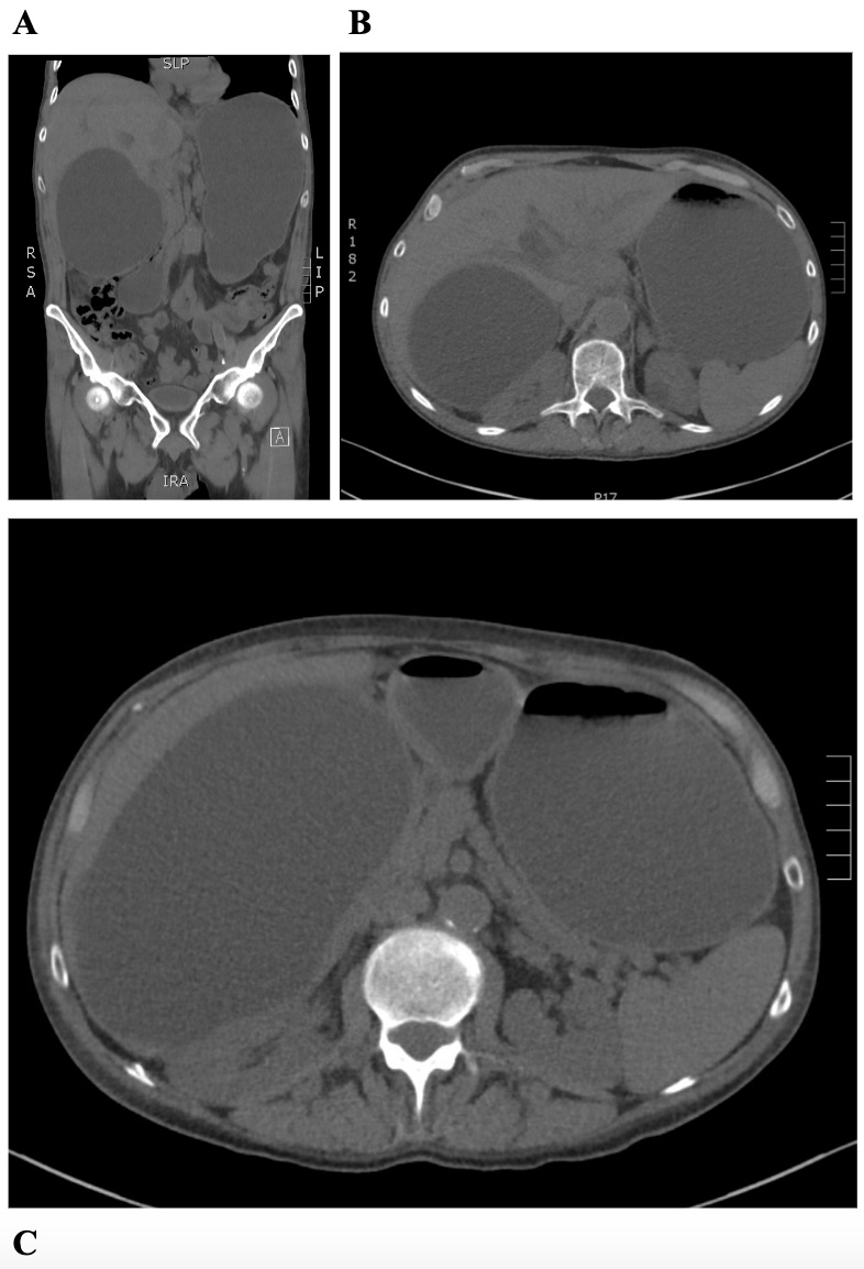
CT Abdomen/Pelvis without contrast revealed a 17.6 cm cystic representing a massively distended gallbladder from new biliary obstruction resulting in GOO with fluid filled, distended stomach and distal esophagus with transition in caliber at proximal duodenum.
A nasogastric tube was placed and IR drainage of the GB, with 1.3L bilious fluid drained. EGD was performed to rule out intra-luminal causes of GOO and revealed slight narrowing in the 2nd portion of the duodenum suggesting extrinsic compression. The patient’s condition overall deteriorated and he unfortunately expired prior to further work up of his cancer.
Discussion: GB mucocele is characterized by GB distension without inflammation, typically due to prolonged cystic duct blockage, in this case malignancy. Our case highlights the importance of considering non-stone related GB mucocele in patients with GOO without a clear cause.

Figure: A: coronal plane showing massively dilated GB and stomach
B: cross-sectional view showing massively dilated GB
C: cross-sectional view showing massively dilated GB
B: cross-sectional view showing massively dilated GB
C: cross-sectional view showing massively dilated GB
Disclosures:
Syed Hamaad Rahman indicated no relevant financial relationships.
Kyle Schneider indicated no relevant financial relationships.
Blake Thompson indicated no relevant financial relationships.
Syed Hamaad Rahman, DO1, Kyle Schneider, MD2, Blake Thompson, MD3. P3544 - The Gargantuan Gallbladder: A Case of Gastric Outlet Obstruction Caused by a Massively Dilated Stoneless Gallbladder, ACG 2024 Annual Scientific Meeting Abstracts. Philadelphia, PA: American College of Gastroenterology.

