Tuesday Poster Session
Category: Biliary/Pancreas
P3553 - Successful Endoscopic Treatment of a Cholecysto-Duodenal Fistula
Tuesday, October 29, 2024
10:30 AM - 4:00 PM ET
Location: Exhibit Hall E

Has Audio
- JM
Jaslyn Maurer, DO
New York-Presbyterian/Queens
Flushing, NY
Presenting Author(s)
Jaslyn Maurer, DO, Gagan Verma, MD, Sang Hoon Kim, MD, Constantine Fisher, MD
New York-Presbyterian/Queens, Flushing, NY
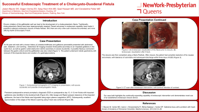
Introduction: Chronic irritation of the gallbladder wall can lead to the development of a cholecystoenteric fistula. Traditionally, cholecystoenteric fistulae have been laparoscopically repaired. Recent advances in endoscopic capability have made it possible to attempt endoscopic closure of these fistulae, however. Still, there are only a few such closures documented, and none utilizing Apollo Endosurgery X-tack. We present a case of an 84-year-old male with a cholecystoenteric fistula repaired endoscopically using X-tack.
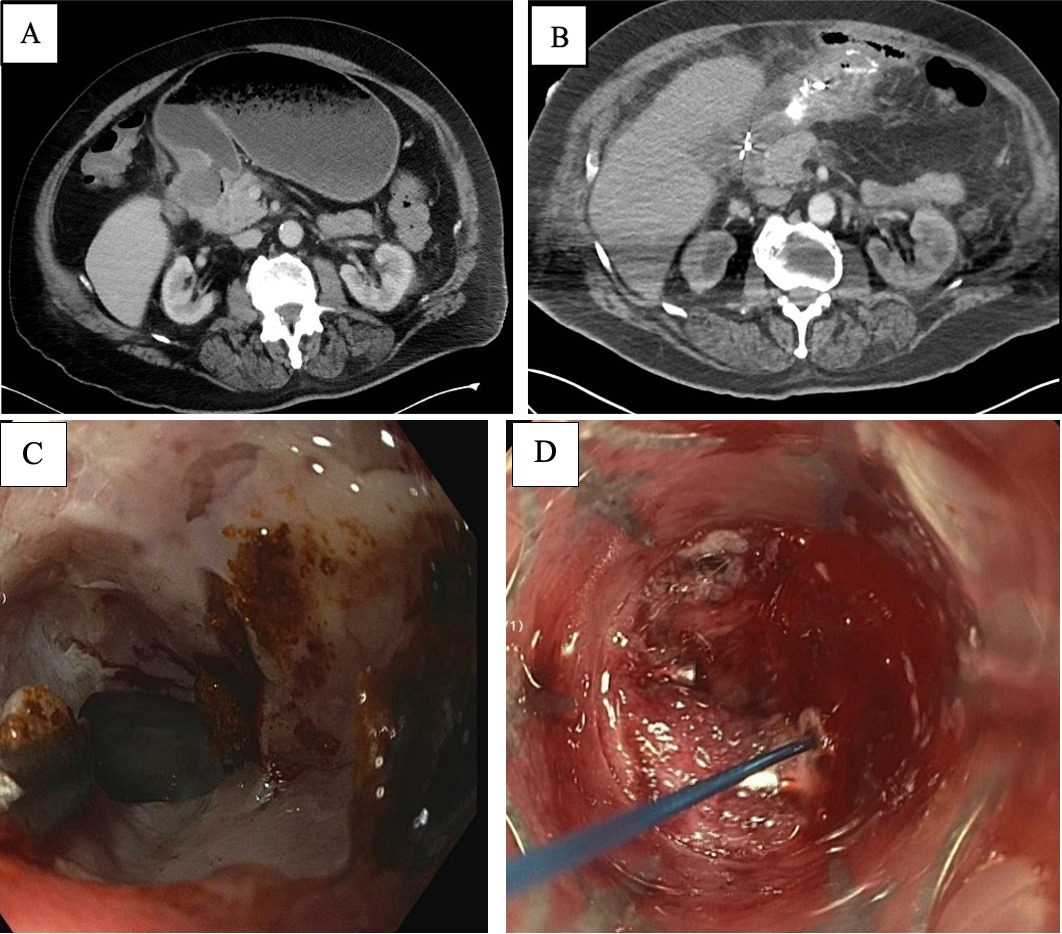
Case Description/Methods: An 84-year-old male with a known history of choledocholithiasis and gallstone pancreatitis presented with abdominal pain, distention, and vomiting. Abdominal 3D imaging revealed cholecystitis secondary to an impacted gallstone in the cystic duct, as well as gastric outlet obstruction (GOO) secondary to severe duodenitis. A possible fistulous connection between the gastric antrum and the gallbladder was also noted (Figure 1A-B). The patient underwent robotic gastrotomy with removal of the impacted stone and creation of a gastrojejunostomy. Persistent post operative emesis prompted a diagnostic EGD on postoperative day 12.
A 12 mm fistula with impacted gallstones was identified in the duodenal bulb (Figure 1C). After lavage and Raptor grasper clearance of the impacted gallstones, the tract of the fistula was fulgurated using argon plasma coagulation (APC). Subsequently, excellent approximation of the edges of the fistula’s opening using X-tack was achieved (Figure 1D). The closure was then completed using a Steris Padlock. After closure, the patient had complete resolution of his nausea and emesis, with tolerance of oral intake and subsequent discharge within three days of EGD.
Discussion: Our case both highlights the continually expanding capability of endoscopic intervention and demonstrates novel use of an established endoscopic closure device.

Disclosures:
Jaslyn Maurer, DO, Gagan Verma, MD, Sang Hoon Kim, MD, Constantine Fisher, MD. P3553 - Successful Endoscopic Treatment of a Cholecysto-Duodenal Fistula, ACG 2024 Annual Scientific Meeting Abstracts. Philadelphia, PA: American College of Gastroenterology.
New York-Presbyterian/Queens, Flushing, NY
Introduction: Chronic irritation of the gallbladder wall can lead to the development of a cholecystoenteric fistula. Traditionally, cholecystoenteric fistulae have been laparoscopically repaired. Recent advances in endoscopic capability have made it possible to attempt endoscopic closure of these fistulae, however. Still, there are only a few such closures documented, and none utilizing Apollo Endosurgery X-tack. We present a case of an 84-year-old male with a cholecystoenteric fistula repaired endoscopically using X-tack.
Case Description/Methods: An 84-year-old male with a known history of choledocholithiasis and gallstone pancreatitis presented with abdominal pain, distention, and vomiting. Abdominal 3D imaging revealed cholecystitis secondary to an impacted gallstone in the cystic duct, as well as gastric outlet obstruction (GOO) secondary to severe duodenitis. A possible fistulous connection between the gastric antrum and the gallbladder was also noted (Figure 1A-B). The patient underwent robotic gastrotomy with removal of the impacted stone and creation of a gastrojejunostomy. Persistent post operative emesis prompted a diagnostic EGD on postoperative day 12.
A 12 mm fistula with impacted gallstones was identified in the duodenal bulb (Figure 1C). After lavage and Raptor grasper clearance of the impacted gallstones, the tract of the fistula was fulgurated using argon plasma coagulation (APC). Subsequently, excellent approximation of the edges of the fistula’s opening using X-tack was achieved (Figure 1D). The closure was then completed using a Steris Padlock. After closure, the patient had complete resolution of his nausea and emesis, with tolerance of oral intake and subsequent discharge within three days of EGD.
Discussion: Our case both highlights the continually expanding capability of endoscopic intervention and demonstrates novel use of an established endoscopic closure device.

Figure: Figure 1: (A) Computerized tomographic (CT) imaging on presentation, with severe duodenitis and possible cholecystogastric fistula. (B) CT scan after endoscopic intervention, with lumenal retention of contrast. (C) Endoscopic visualization of the fistula prior to closure. (D) Endoscopic visualization after X-tack.
Disclosures:
Jaslyn Maurer indicated no relevant financial relationships.
Gagan Verma indicated no relevant financial relationships.
Sang Hoon Kim indicated no relevant financial relationships.
Constantine Fisher indicated no relevant financial relationships.
Jaslyn Maurer, DO, Gagan Verma, MD, Sang Hoon Kim, MD, Constantine Fisher, MD. P3553 - Successful Endoscopic Treatment of a Cholecysto-Duodenal Fistula, ACG 2024 Annual Scientific Meeting Abstracts. Philadelphia, PA: American College of Gastroenterology.
