Tuesday Poster Session
Category: Biliary/Pancreas
P3615 - A Rare Ca(u)se of Metastatic Renal Cell Carcinoma in the Biliary Tree
Tuesday, October 29, 2024
10:30 AM - 4:00 PM ET
Location: Exhibit Hall E

Has Audio
- SP
Sailaja Pisipati, MBBS
Mayo Clinic
Atlanta, GA
Presenting Author(s)
Award: Presidential Poster Award
Sailaja Pisipati, MBBS, Sumera Ilyas, MD, Aliana Bofill-Garcia, MD
Mayo Clinic, Rochester, MN
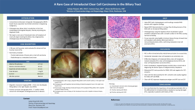
Introduction: Conventional endoscopic retrograde cholangiography (ERCP) based tissue acquisition has limited sensitivity in diagnosing malignancy. Cholangioscopy allows direct visualization of stricture morphology and targeted biopsies, thereby improving the diagnostic yield. We report a rare case of intraductal clear cell neoplasm of renal cell origin involving the biliary tree, diagnosed by cholangioscopy-directed biopsies.
Case Description/Methods: A 68-year-old Caucasian man was evaluated for abnormal liver function tests. He underwent a partial nephrectomy 10 years prior for renal cell carcinoma, and radiotherapy to a metastatic bony lesion. Alkaline phosphatase was 177 U/L, alanine aminotransferase 81 U/L, aspartate aminotransferase 61 U/L, and total bilirubin 1 mg/dl. Magnetic resonance imaging demonstrated a 17x11mm intrahepatic filling defect in the right hepatic duct. Positron emission tomography showed increased avidity in this area, raising concern for intrahepatic cholangiocarcinoma versus abscess. Ca 19-9 was 393 U/ml, alpha fetoprotein was normal. Index ERCP with brushings revealed right hepatic duct stenosis with negative cytology. Repeat ERCP demonstrated 2 cm long, flow-limiting stenosis of the right main hepatic duct with an intraductal filling defect. Cholangioscopy using the SpyGlass direct visualization system revealed a polypoid mass with a smooth surface in the right hepatic duct, causing luminal narrowing. Tissue acquired using SpyBite miniature biopsy forceps showed clear cell neoplasm with positive PAX-8 and CAIX on immunohistochemistry (IHC), confirming metastatic renal cell carcinoma (RCC).
Discussion: RCC is often characterized by metachronous lesions to unusual sites. Intraductal metastatic clear cell neoplasms are extremely rare. Differential diagnosis of intraductal biliary clear cell neoplasms include metastatic RCC, clear cell cholangiocarcinoma (CCA), and clear cell hepatocellular carcinoma (HCC). Direct visualization, targeted biopsies and IHC play a crucial role in diagnosing the diverse types of malignant biliary neoplasms. Metastatic RCC stains positive for PAX8, CAIX and negative for CK7, vimentin. Clear cell CCA stains positive for CK7, vimentin and usually negative for CK20, AFP and PAX8. Clear cell HCC expresses hepatocyte antigen and AFP, but negative for CK7. Our case illustrates the importance of considering metastatic RCC in the differentials for intraductal biliary neoplasms.

Disclosures:
Sailaja Pisipati, MBBS, Sumera Ilyas, MD, Aliana Bofill-Garcia, MD. P3615 - A Rare Ca(u)se of Metastatic Renal Cell Carcinoma in the Biliary Tree, ACG 2024 Annual Scientific Meeting Abstracts. Philadelphia, PA: American College of Gastroenterology.
Sailaja Pisipati, MBBS, Sumera Ilyas, MD, Aliana Bofill-Garcia, MD
Mayo Clinic, Rochester, MN
Introduction: Conventional endoscopic retrograde cholangiography (ERCP) based tissue acquisition has limited sensitivity in diagnosing malignancy. Cholangioscopy allows direct visualization of stricture morphology and targeted biopsies, thereby improving the diagnostic yield. We report a rare case of intraductal clear cell neoplasm of renal cell origin involving the biliary tree, diagnosed by cholangioscopy-directed biopsies.
Case Description/Methods: A 68-year-old Caucasian man was evaluated for abnormal liver function tests. He underwent a partial nephrectomy 10 years prior for renal cell carcinoma, and radiotherapy to a metastatic bony lesion. Alkaline phosphatase was 177 U/L, alanine aminotransferase 81 U/L, aspartate aminotransferase 61 U/L, and total bilirubin 1 mg/dl. Magnetic resonance imaging demonstrated a 17x11mm intrahepatic filling defect in the right hepatic duct. Positron emission tomography showed increased avidity in this area, raising concern for intrahepatic cholangiocarcinoma versus abscess. Ca 19-9 was 393 U/ml, alpha fetoprotein was normal. Index ERCP with brushings revealed right hepatic duct stenosis with negative cytology. Repeat ERCP demonstrated 2 cm long, flow-limiting stenosis of the right main hepatic duct with an intraductal filling defect. Cholangioscopy using the SpyGlass direct visualization system revealed a polypoid mass with a smooth surface in the right hepatic duct, causing luminal narrowing. Tissue acquired using SpyBite miniature biopsy forceps showed clear cell neoplasm with positive PAX-8 and CAIX on immunohistochemistry (IHC), confirming metastatic renal cell carcinoma (RCC).
Discussion: RCC is often characterized by metachronous lesions to unusual sites. Intraductal metastatic clear cell neoplasms are extremely rare. Differential diagnosis of intraductal biliary clear cell neoplasms include metastatic RCC, clear cell cholangiocarcinoma (CCA), and clear cell hepatocellular carcinoma (HCC). Direct visualization, targeted biopsies and IHC play a crucial role in diagnosing the diverse types of malignant biliary neoplasms. Metastatic RCC stains positive for PAX8, CAIX and negative for CK7, vimentin. Clear cell CCA stains positive for CK7, vimentin and usually negative for CK20, AFP and PAX8. Clear cell HCC expresses hepatocyte antigen and AFP, but negative for CK7. Our case illustrates the importance of considering metastatic RCC in the differentials for intraductal biliary neoplasms.

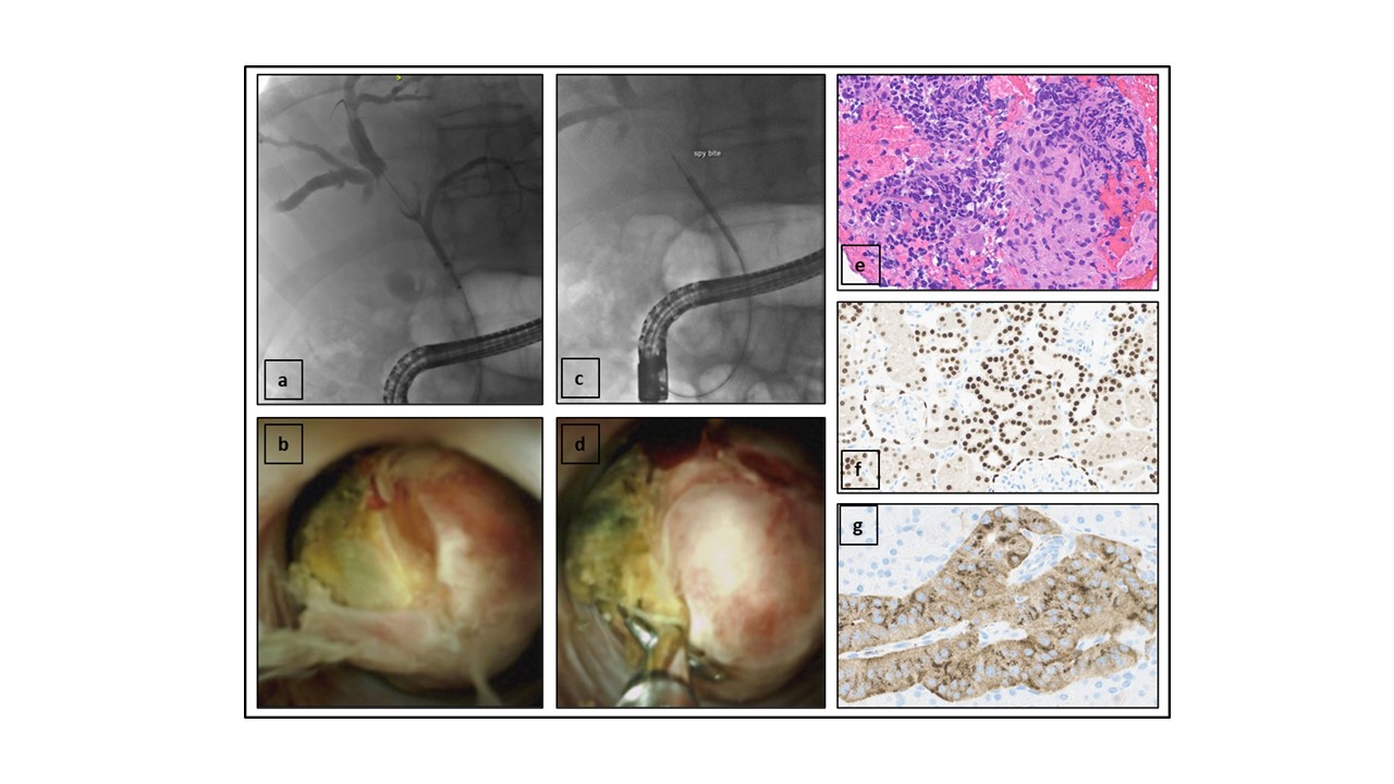
Figure: (a) Polypoid filling defect with smooth outline, in the right main hepatic duct on cholangiography
(b) Cholangioscopy demonstrating the intraductal polypoid mass in right hepatic duct
(c) Fluoroscopic image showing intraductal biopsy with spybite miniature biopsy forceps
(d) Cholangioscopic view of biopsy of the intraductal mass with spybite miniature biopsy forceps
(e) Hematoxylin and eosin stain showing glycogen-laden clear cell neoplasm
(f) Positive staining with PAX-8
(g) Positive staining with CAIX
Disclosures:
Sailaja Pisipati indicated no relevant financial relationships.
Sumera Ilyas indicated no relevant financial relationships.
Aliana Bofill-Garcia indicated no relevant financial relationships.
Sailaja Pisipati, MBBS, Sumera Ilyas, MD, Aliana Bofill-Garcia, MD. P3615 - A Rare Ca(u)se of Metastatic Renal Cell Carcinoma in the Biliary Tree, ACG 2024 Annual Scientific Meeting Abstracts. Philadelphia, PA: American College of Gastroenterology.

