Tuesday Poster Session
Category: Colon
P3732 - Nerve-Wracking but Not Gut-Wrenching: A Rare Case of a Colonic Ganglioneuroma
Tuesday, October 29, 2024
10:30 AM - 4:00 PM ET
Location: Exhibit Hall E

Has Audio
- PS
Priyanka Shah, MD
Lewis Katz School of Medicine at Temple University
Philadelphia, PA
Presenting Author(s)
Priyanka Shah, MD, Frank Friedenberg, MD, MS
Lewis Katz School of Medicine at Temple University, Philadelphia, PA
Introduction: Colonic ganglioneuromas (CGN) are rare, benign tumors that may cause abdominal pain, weight loss, constipation, bleeding, and/or obstruction, but also may present asymptomatically. CGN is often positive for S-100 and sometimes positive for synaptophysin. We present the case of a CGN that was negative for S-100 underneath a sessile serrated lesion (SSL).
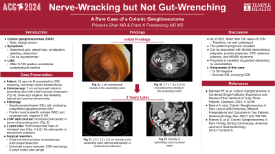
Case Description/Methods: A 50-year-old male presented to clinic for colorectal cancer screening. Other than small-volume hematochezia with defecation, he was asymptomatic. Colonoscopy revealed not only non-bleeding internal and external hemorrhoids, the source of his hematochezia, but also a 20 mm submucosal nodule in the ascending colon (Fig. A). Pathology showed an SSL with an underlying subepithelial ganglioneuroma (GN) that was positive for neuron-specific enolase (NSE) and synaptophysin but negative for S-100. A follow-up CT abdomen/pelvis with contrast further characterized the GN: a 2.7 x 1.8 x 3.0 cm nonobstructive density in the ascending colon’s lumen near the hepatic flexure (Fig. B). 3 years later, repeat CT showed interval enlargement of the GN to 2.6 x 2.5 x 2.5 cm without adenopathy or extracolonic extension (Fig. C). The patient was referred to colorectal surgery as removal of the lesion was not amenable to endoscopic submucosal dissection.
Discussion: As of 2023, fewer than 100 cases of CGN have been described. Based on existing literature, this patient’s polypoid CGN should be resected and his prognosis should be excellent. However, other types of CGN can be associated with diseases such as MEN2b syndrome, so in those patients, prognosis is guarded. Two factors differentiated this case from many others in the literature: the patient’s CGN was S-100 negative and there was a mucosal SSL overlying the lesion. This case demonstrates that there remains significant variation in the findings of CGNs that are not well-understood.
Bahmad HF, et al. Colonic Ganglioneuroma: A Combined Single-Institution Experience and Review of the Literature of Forty-Three Patients. Diseases. 2023; 11(2):69.
Baiomi A, et al. Colonic Ganglioneuroma: A Rare Lesion With Extremely Different Presentations and Outcomes in Two Patients. Gastroenterology Res. 2021;14(3):194-198.
Etienne D, et al. Colonic Ganglioneuroma: A Rare Finding During Colonoscopy. American Journal of Gastroenterology. 2016;111:618-619.

Disclosures:
Priyanka Shah, MD, Frank Friedenberg, MD, MS. P3732 - Nerve-Wracking but Not Gut-Wrenching: A Rare Case of a Colonic Ganglioneuroma, ACG 2024 Annual Scientific Meeting Abstracts. Philadelphia, PA: American College of Gastroenterology.
Lewis Katz School of Medicine at Temple University, Philadelphia, PA
Introduction: Colonic ganglioneuromas (CGN) are rare, benign tumors that may cause abdominal pain, weight loss, constipation, bleeding, and/or obstruction, but also may present asymptomatically. CGN is often positive for S-100 and sometimes positive for synaptophysin. We present the case of a CGN that was negative for S-100 underneath a sessile serrated lesion (SSL).
Case Description/Methods: A 50-year-old male presented to clinic for colorectal cancer screening. Other than small-volume hematochezia with defecation, he was asymptomatic. Colonoscopy revealed not only non-bleeding internal and external hemorrhoids, the source of his hematochezia, but also a 20 mm submucosal nodule in the ascending colon (Fig. A). Pathology showed an SSL with an underlying subepithelial ganglioneuroma (GN) that was positive for neuron-specific enolase (NSE) and synaptophysin but negative for S-100. A follow-up CT abdomen/pelvis with contrast further characterized the GN: a 2.7 x 1.8 x 3.0 cm nonobstructive density in the ascending colon’s lumen near the hepatic flexure (Fig. B). 3 years later, repeat CT showed interval enlargement of the GN to 2.6 x 2.5 x 2.5 cm without adenopathy or extracolonic extension (Fig. C). The patient was referred to colorectal surgery as removal of the lesion was not amenable to endoscopic submucosal dissection.
Discussion: As of 2023, fewer than 100 cases of CGN have been described. Based on existing literature, this patient’s polypoid CGN should be resected and his prognosis should be excellent. However, other types of CGN can be associated with diseases such as MEN2b syndrome, so in those patients, prognosis is guarded. Two factors differentiated this case from many others in the literature: the patient’s CGN was S-100 negative and there was a mucosal SSL overlying the lesion. This case demonstrates that there remains significant variation in the findings of CGNs that are not well-understood.
Bahmad HF, et al. Colonic Ganglioneuroma: A Combined Single-Institution Experience and Review of the Literature of Forty-Three Patients. Diseases. 2023; 11(2):69.
Baiomi A, et al. Colonic Ganglioneuroma: A Rare Lesion With Extremely Different Presentations and Outcomes in Two Patients. Gastroenterology Res. 2021;14(3):194-198.
Etienne D, et al. Colonic Ganglioneuroma: A Rare Finding During Colonoscopy. American Journal of Gastroenterology. 2016;111:618-619.

Figure: A: 20 mm submucosal nodule in the ascending colon on colonoscopy
B: 2.7 x 1.8 x 3.0 cm nonobstructive density in the ascending colon on CT
C: 2.6 x 2.5 x 2.5 cm density in the ascending colon without adenopathy or extracolonic extension on CT (3 years after Fig. B)
B: 2.7 x 1.8 x 3.0 cm nonobstructive density in the ascending colon on CT
C: 2.6 x 2.5 x 2.5 cm density in the ascending colon without adenopathy or extracolonic extension on CT (3 years after Fig. B)
Disclosures:
Priyanka Shah indicated no relevant financial relationships.
Frank Friedenberg indicated no relevant financial relationships.
Priyanka Shah, MD, Frank Friedenberg, MD, MS. P3732 - Nerve-Wracking but Not Gut-Wrenching: A Rare Case of a Colonic Ganglioneuroma, ACG 2024 Annual Scientific Meeting Abstracts. Philadelphia, PA: American College of Gastroenterology.
