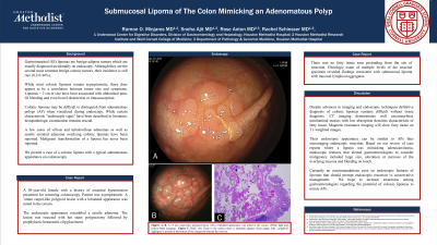
Tuesday Poster Session
Category: Colon
P3733 - Submucosal Lipoma of the Colon Mimicking an Adenomatous Polyp
Tuesday, October 29, 2024
10:30 AM - 4:00 PM ET
Location: Exhibit Hall E

Has Audio
- RM
Ramon O. Minjares, MD
Underwood Center for Digestive Disorders, Houston Methodist Hospital
Houston, TX
Presenting Author(s)
Ramon O.. Minjares, MD1, Sneha Ajit, MD2, Rose Anton, MD2, Rachel Schiesser, MD1
1Underwood Center for Digestive Disorders, Houston Methodist Hospital, Houston, TX; 2Houston Methodist Hospital, Houston, TX
Introduction: Gastrointestinal (GI) lipomas are benign adipose tumors which are usually diagnosed incidentally on endoscopy. Although they are the second most common benign colonic tumors, their incidence is still rare (0.2-0.44%). While most colonic lipomas remain asymptomatic, there does appear to be a correlation between tumor size and symptoms. Lipomas > 2 cm in size have been associated with abdominal pain, GI bleeding and even bowel obstruction or intussusception. Colonic lipomas may be difficult to distinguish from adenomatous polyps (AP) when visualized during endoscopy. While certain characteristic “endoscopic signs” have been described in literature, histopathologic examination remains crucial. A few cases of villous and tubulovillous adenomas as well as sessile serrated adenoma overlying colonic lipomas have been reported. Malignant transformation of a lipoma has never been reported. We present a case of a colonic lipoma with a typical adenomatous appearance on colonoscopy.
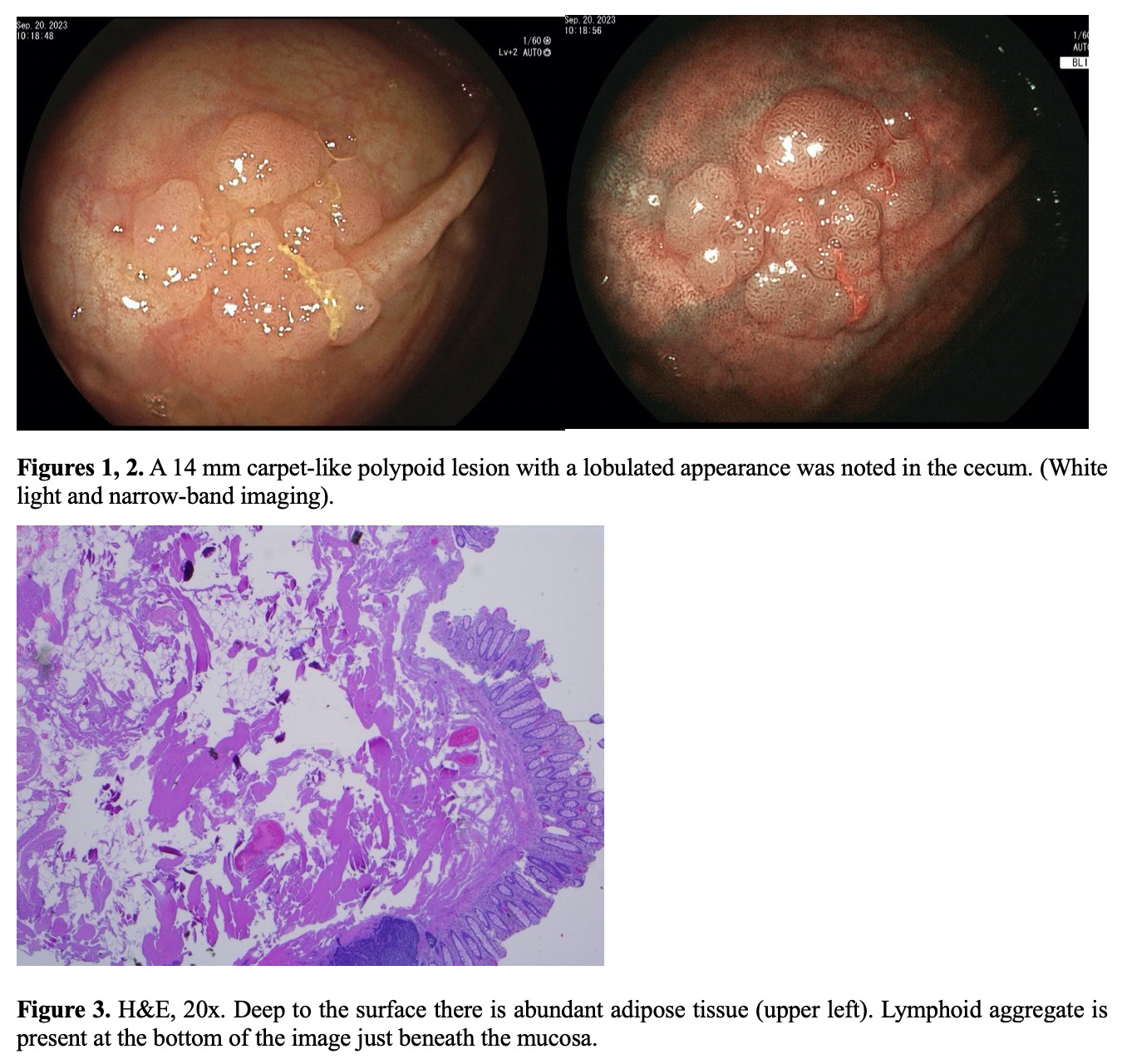
Case Description/Methods: A 50-year-old female with history of hypertension presented for screening colonoscopy. Patient was asymptomatic. A 14mm carpet-like polypoid lesion with a lobulated appearance was noted in the cecum. The endoscopic appearance resembled a sessile adenoma. The lesion was resected with hot snare polypectomy followed by prophylactic hemostatic clip placement. There was no fatty tissue seen to be protruding from the site of resection. Histologic exam of multiple levels of the resected specimen revealed findings consistent with submucosal lipoma with mucosal lymphoid aggregates.
Discussion: Despite advances in imaging and endoscopic techniques definitive diagnosis of colonic lipomas remains difficult without tissue diagnosis. CT imaging demonstrates well circumscribed, intraluminal masses with low absorption densities characteristic of fatty tissue. Magnetic resonance imaging will show fatty tissue on T1-weighted images. Their endoscopic appearance can be similar to APs thus encouraging endoscopic resection. Based on our review of case reports where a lipoma was mimicking adenocarcinoma, endoscopic features that alerted gastroenterologists to consider malignancy included large size, ulceration or necrosis of the overlying mucosa and bleeding on touch. Currently no recommendations exist on endoscopic features of lipomas that should prompt endoscopic resection vs conservative management. We hope to increase awareness among gastroenterologists regarding the potential of colonic lipomas to mimic APs.

Disclosures:
Ramon O.. Minjares, MD1, Sneha Ajit, MD2, Rose Anton, MD2, Rachel Schiesser, MD1. P3733 - Submucosal Lipoma of the Colon Mimicking an Adenomatous Polyp, ACG 2024 Annual Scientific Meeting Abstracts. Philadelphia, PA: American College of Gastroenterology.
1Underwood Center for Digestive Disorders, Houston Methodist Hospital, Houston, TX; 2Houston Methodist Hospital, Houston, TX
Introduction: Gastrointestinal (GI) lipomas are benign adipose tumors which are usually diagnosed incidentally on endoscopy. Although they are the second most common benign colonic tumors, their incidence is still rare (0.2-0.44%). While most colonic lipomas remain asymptomatic, there does appear to be a correlation between tumor size and symptoms. Lipomas > 2 cm in size have been associated with abdominal pain, GI bleeding and even bowel obstruction or intussusception. Colonic lipomas may be difficult to distinguish from adenomatous polyps (AP) when visualized during endoscopy. While certain characteristic “endoscopic signs” have been described in literature, histopathologic examination remains crucial. A few cases of villous and tubulovillous adenomas as well as sessile serrated adenoma overlying colonic lipomas have been reported. Malignant transformation of a lipoma has never been reported. We present a case of a colonic lipoma with a typical adenomatous appearance on colonoscopy.
Case Description/Methods: A 50-year-old female with history of hypertension presented for screening colonoscopy. Patient was asymptomatic. A 14mm carpet-like polypoid lesion with a lobulated appearance was noted in the cecum. The endoscopic appearance resembled a sessile adenoma. The lesion was resected with hot snare polypectomy followed by prophylactic hemostatic clip placement. There was no fatty tissue seen to be protruding from the site of resection. Histologic exam of multiple levels of the resected specimen revealed findings consistent with submucosal lipoma with mucosal lymphoid aggregates.
Discussion: Despite advances in imaging and endoscopic techniques definitive diagnosis of colonic lipomas remains difficult without tissue diagnosis. CT imaging demonstrates well circumscribed, intraluminal masses with low absorption densities characteristic of fatty tissue. Magnetic resonance imaging will show fatty tissue on T1-weighted images. Their endoscopic appearance can be similar to APs thus encouraging endoscopic resection. Based on our review of case reports where a lipoma was mimicking adenocarcinoma, endoscopic features that alerted gastroenterologists to consider malignancy included large size, ulceration or necrosis of the overlying mucosa and bleeding on touch. Currently no recommendations exist on endoscopic features of lipomas that should prompt endoscopic resection vs conservative management. We hope to increase awareness among gastroenterologists regarding the potential of colonic lipomas to mimic APs.

Figure: Figures 1, 2. A 14 mm carpet-like polypoid lesion with a lobulated appearance was noted in the cecum. (White light and narrow-band imaging).
Figure 3. H&E, 20x. Deep to the surface there is abundant adipose tissue (upper left). Lymphoid aggregate is present at the bottom of the image just beneath the mucosa.
Figure 3. H&E, 20x. Deep to the surface there is abundant adipose tissue (upper left). Lymphoid aggregate is present at the bottom of the image just beneath the mucosa.
Disclosures:
Ramon Minjares indicated no relevant financial relationships.
Sneha Ajit indicated no relevant financial relationships.
Rose Anton indicated no relevant financial relationships.
Rachel Schiesser indicated no relevant financial relationships.
Ramon O.. Minjares, MD1, Sneha Ajit, MD2, Rose Anton, MD2, Rachel Schiesser, MD1. P3733 - Submucosal Lipoma of the Colon Mimicking an Adenomatous Polyp, ACG 2024 Annual Scientific Meeting Abstracts. Philadelphia, PA: American College of Gastroenterology.
