Tuesday Poster Session
Category: Colon
P3736 - Barium Contrast Induced Bowel Obstruction Treated With Endoscopic Retrieval
Tuesday, October 29, 2024
10:30 AM - 4:00 PM ET
Location: Exhibit Hall E

Has Audio

Robert Tamai, MD
David Geffen School of Medicine at UCLA
Los Angeles, CA
Presenting Author(s)
Award: Presidential Poster Award
Robert Tamai, MD, Selena Zhou, MD, Robert Mocharla, MD
David Geffen School of Medicine at UCLA, Los Angeles, CA
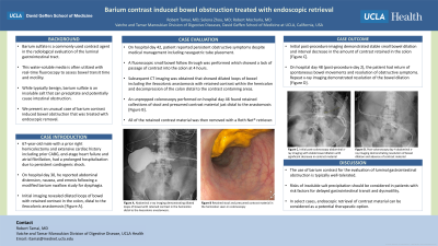
Introduction: Barium sulfate is a commonly used contrast agent in the radiological evaluation of the luminal gastrointestinal tract. This water-soluble media is often utilized with real-time fluoroscopy to assess bowel transit time and motility. While typically benign, barium sulfate is an insoluble salt that can precipitate and potentially cause intestinal obstruction. We present an unusual case of barium contrast induced bowel obstruction that was treated with endoscopic retrieval.
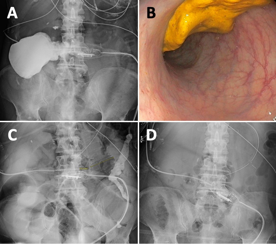
Case Description/Methods: A 67-year-old male with a prior right hemicolectomy and extensive cardiac history including prior CABG, end-stage heart failure, and atrial fibrillation, had a prolonged hospitalization due to persistent cardiogenic shock. On hospital day 30, he reported abdominal distension, nausea, and emesis following a modified barium swallow study for dysphagia. Initial imaging revealed dilated loops of bowel with retained contrast in the colon, distal to the ileocolonic anastomosis [Figure A].
On hospital day 42, patient reported persistent obstructive symptoms despite medical management including nasogastric tube placement. A fluoroscopic small bowel follow through was performed which showed a lack of passage of contrast into the colon at 4 hours. Subsequent CT imaging was obtained that showed dilated loops of bowel including the ileocolonic anastomosis with retained contrast within the hemicolon and decompression of the colon distal to the contrast containing areas.
An unprepped colonoscopy performed on hospital day 46 found retained collections of stool and presumed contrast material just distal to the anastomosis [Figure B]. All of the retained contrast material was then removed with a Roth Net® retriever. Initial post-procedure imaging demonstrated stable small bowel dilation and interval decrease in the amount of contrast retained in the colon [Figure C]. On hospital day 48 (post-procedure day 2), the patient had return of spontaneous bowel movements, resolution of obstructive symptoms, and repeat x-ray imaging demonstrated resolution of the bowel dilation [Figure D].
Discussion: The use of barium contrast for the evaluation of luminal gastrointestinal obstruction is typically well-tolerated. However, the risks of insoluble salt precipitation should be considered, especially in patients with risk factors for delayed gastrointestinal transit and dysmotility. In select cases, endoscopic retrieval of contrast material can be considered as a potential therapeutic option.

Disclosures:
Robert Tamai, MD, Selena Zhou, MD, Robert Mocharla, MD. P3736 - Barium Contrast Induced Bowel Obstruction Treated With Endoscopic Retrieval, ACG 2024 Annual Scientific Meeting Abstracts. Philadelphia, PA: American College of Gastroenterology.
Robert Tamai, MD, Selena Zhou, MD, Robert Mocharla, MD
David Geffen School of Medicine at UCLA, Los Angeles, CA
Introduction: Barium sulfate is a commonly used contrast agent in the radiological evaluation of the luminal gastrointestinal tract. This water-soluble media is often utilized with real-time fluoroscopy to assess bowel transit time and motility. While typically benign, barium sulfate is an insoluble salt that can precipitate and potentially cause intestinal obstruction. We present an unusual case of barium contrast induced bowel obstruction that was treated with endoscopic retrieval.
Case Description/Methods: A 67-year-old male with a prior right hemicolectomy and extensive cardiac history including prior CABG, end-stage heart failure, and atrial fibrillation, had a prolonged hospitalization due to persistent cardiogenic shock. On hospital day 30, he reported abdominal distension, nausea, and emesis following a modified barium swallow study for dysphagia. Initial imaging revealed dilated loops of bowel with retained contrast in the colon, distal to the ileocolonic anastomosis [Figure A].
On hospital day 42, patient reported persistent obstructive symptoms despite medical management including nasogastric tube placement. A fluoroscopic small bowel follow through was performed which showed a lack of passage of contrast into the colon at 4 hours. Subsequent CT imaging was obtained that showed dilated loops of bowel including the ileocolonic anastomosis with retained contrast within the hemicolon and decompression of the colon distal to the contrast containing areas.
An unprepped colonoscopy performed on hospital day 46 found retained collections of stool and presumed contrast material just distal to the anastomosis [Figure B]. All of the retained contrast material was then removed with a Roth Net® retriever. Initial post-procedure imaging demonstrated stable small bowel dilation and interval decrease in the amount of contrast retained in the colon [Figure C]. On hospital day 48 (post-procedure day 2), the patient had return of spontaneous bowel movements, resolution of obstructive symptoms, and repeat x-ray imaging demonstrated resolution of the bowel dilation [Figure D].
Discussion: The use of barium contrast for the evaluation of luminal gastrointestinal obstruction is typically well-tolerated. However, the risks of insoluble salt precipitation should be considered, especially in patients with risk factors for delayed gastrointestinal transit and dysmotility. In select cases, endoscopic retrieval of contrast material can be considered as a potential therapeutic option.

Figure: Figure A: Abdominal x-ray imaging demonstrating dilated loops of bowel with retained contrast in the hemicolon distal to the ileocolonic anastomosis.
Figure B: Retained stool and presumed contrast material in the hemicolon seen on colonoscopy
Figure C: Initial post-colonoscopy abdominal x-ray imaging demonstrating stable bowel dilation, no evidence of pneumatosis or free air, with significant interval decrease in contrast material
Figure D: Post-colonoscopy day 4 abdominal x-ray imaging demonstrating resolution of bowel dilation and absence of contrast material.
Disclosures:
Robert Tamai indicated no relevant financial relationships.
Selena Zhou indicated no relevant financial relationships.
Robert Mocharla indicated no relevant financial relationships.
Robert Tamai, MD, Selena Zhou, MD, Robert Mocharla, MD. P3736 - Barium Contrast Induced Bowel Obstruction Treated With Endoscopic Retrieval, ACG 2024 Annual Scientific Meeting Abstracts. Philadelphia, PA: American College of Gastroenterology.

