Tuesday Poster Session
Category: Colon
P3751 - Unraveling Recurrent Acute Appendicitis: Atypical Presentations and Clinical Considerations
Tuesday, October 29, 2024
10:30 AM - 4:00 PM ET
Location: Exhibit Hall E


Muhammad Jahanzaib Khan, MD
Mather Hospital, Northwell Health
New York, NY
Presenting Author(s)
Abdul Wasaay Khurram, MBBS1, Muhammad Abdurrahman Butt, MBBS1, Muhammad Shahbaz, MBBS1, Muhammad Jahanzaib Khan, MD2, Khurram Qadir, MD3
1Shifa College of Medicine, Islamabad, Islamabad, Pakistan; 2Mather Hospital, Northwell Health, New York, NY; 3Feinberg School of Medicine, Northwestern University, Darien, IL
Introduction: Acute appendicitis is a prevalent cause of acute abdomen and constitutes an urgent abdominal emergency. Traditionally, it is viewed as a progressive condition potentially leading to gangrene and perforation, thus necessitating prompt diagnosis and intervention. However, approximately 10% of patients experience recurrent acute appendicitis, characterized by episodes of right lower quadrant (RLQ) pain that remits and recurs, resolving completely post-appendectomy.
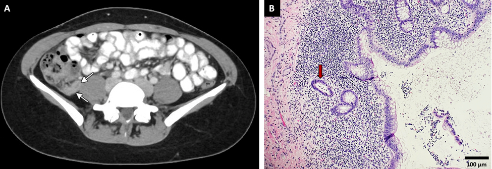
Case Description/Methods: A 22-year-old female with a history of multiple food allergies presented with episodes of intermittent abdominal pain over the past four years. Initially infrequent, the episodes started occurring monthly in the last six months. The sharp periumbilical pain typically commenced two hours post-dinner, lasting around six hours. Recent episodes were accompanied by severe nausea and vomiting, which temporarily alleviated the pain. A computed tomography (CT) scan of the abdomen revealed a retrocecal appendix with a thickened, hyper-enhancing wall and peri-appendiceal fat stranding, indicative of acute appendicitis. Following an appendectomy, histopathology confirmed acute inflammation with focal intraepithelial neutrophils and intraluminal neutrophils in crypts. The patient’s postoperative course was uneventful, and she remained symptom-free at the 12-month follow-up, confirming a diagnosis of recurrent acute appendicitis.
Discussion: Recurrent acute appendicitis is marked by recurrent RLQ pain resolving after appendectomy but can be mistaken for chronic appendicitis, which involves continuous RLQ pain and histological findings of lymphocytes, eosinophils, and fibrosis in the appendiceal wall. Our case involved recurrent acute appendicitis with non-migratory, non-radiating periumbilical rather than RLQ pain. The pain was linked to food intake and accompanied by severe vomiting which provided transient relief. These features are rarely reported in literature. Recurrent appendicitis may result from intermittent appendiceal lumen obstruction or primary torsion and detorsion of the appendix. Elevated tryptase levels in acute appendicitis suggest an allergic component, which is relevant given our patient's multiple food allergies. Diagnosing recurrent appendicitis is challenging, especially in females, where symptoms can mimic gynecological conditions like pelvic inflammatory disease. Missed diagnoses can lead to severe complications such as perforation, abscess, and peritonitis. Definitive management is appendectomy.

Disclosures:
Abdul Wasaay Khurram, MBBS1, Muhammad Abdurrahman Butt, MBBS1, Muhammad Shahbaz, MBBS1, Muhammad Jahanzaib Khan, MD2, Khurram Qadir, MD3. P3751 - Unraveling Recurrent Acute Appendicitis: Atypical Presentations and Clinical Considerations, ACG 2024 Annual Scientific Meeting Abstracts. Philadelphia, PA: American College of Gastroenterology.
1Shifa College of Medicine, Islamabad, Islamabad, Pakistan; 2Mather Hospital, Northwell Health, New York, NY; 3Feinberg School of Medicine, Northwestern University, Darien, IL
Introduction: Acute appendicitis is a prevalent cause of acute abdomen and constitutes an urgent abdominal emergency. Traditionally, it is viewed as a progressive condition potentially leading to gangrene and perforation, thus necessitating prompt diagnosis and intervention. However, approximately 10% of patients experience recurrent acute appendicitis, characterized by episodes of right lower quadrant (RLQ) pain that remits and recurs, resolving completely post-appendectomy.
Case Description/Methods: A 22-year-old female with a history of multiple food allergies presented with episodes of intermittent abdominal pain over the past four years. Initially infrequent, the episodes started occurring monthly in the last six months. The sharp periumbilical pain typically commenced two hours post-dinner, lasting around six hours. Recent episodes were accompanied by severe nausea and vomiting, which temporarily alleviated the pain. A computed tomography (CT) scan of the abdomen revealed a retrocecal appendix with a thickened, hyper-enhancing wall and peri-appendiceal fat stranding, indicative of acute appendicitis. Following an appendectomy, histopathology confirmed acute inflammation with focal intraepithelial neutrophils and intraluminal neutrophils in crypts. The patient’s postoperative course was uneventful, and she remained symptom-free at the 12-month follow-up, confirming a diagnosis of recurrent acute appendicitis.
Discussion: Recurrent acute appendicitis is marked by recurrent RLQ pain resolving after appendectomy but can be mistaken for chronic appendicitis, which involves continuous RLQ pain and histological findings of lymphocytes, eosinophils, and fibrosis in the appendiceal wall. Our case involved recurrent acute appendicitis with non-migratory, non-radiating periumbilical rather than RLQ pain. The pain was linked to food intake and accompanied by severe vomiting which provided transient relief. These features are rarely reported in literature. Recurrent appendicitis may result from intermittent appendiceal lumen obstruction or primary torsion and detorsion of the appendix. Elevated tryptase levels in acute appendicitis suggest an allergic component, which is relevant given our patient's multiple food allergies. Diagnosing recurrent appendicitis is challenging, especially in females, where symptoms can mimic gynecological conditions like pelvic inflammatory disease. Missed diagnoses can lead to severe complications such as perforation, abscess, and peritonitis. Definitive management is appendectomy.

Figure: A. Contrast enhanced CT scan showing a retrocecal appendix with a thickened and hyper-enhancing wall, indicated by white arrows.
B. Histologic section of the appendix at low power shows colonic mucosa with lymphoid tissue in the mucosa and submucosa. Intraluminal neutrophils in a crypt are indicated by the red arrow, representing acute inflammation.
B. Histologic section of the appendix at low power shows colonic mucosa with lymphoid tissue in the mucosa and submucosa. Intraluminal neutrophils in a crypt are indicated by the red arrow, representing acute inflammation.
Disclosures:
Abdul Wasaay Khurram indicated no relevant financial relationships.
Muhammad Abdurrahman Butt indicated no relevant financial relationships.
Muhammad Shahbaz indicated no relevant financial relationships.
Muhammad Jahanzaib Khan indicated no relevant financial relationships.
Khurram Qadir indicated no relevant financial relationships.
Abdul Wasaay Khurram, MBBS1, Muhammad Abdurrahman Butt, MBBS1, Muhammad Shahbaz, MBBS1, Muhammad Jahanzaib Khan, MD2, Khurram Qadir, MD3. P3751 - Unraveling Recurrent Acute Appendicitis: Atypical Presentations and Clinical Considerations, ACG 2024 Annual Scientific Meeting Abstracts. Philadelphia, PA: American College of Gastroenterology.
