Tuesday Poster Session
Category: Colon
P3763 - A Case of a Type IV Hiatal Hernia Presenting With Sinus Bradycardia
Tuesday, October 29, 2024
10:30 AM - 4:00 PM ET
Location: Exhibit Hall E

Has Audio

Sahil Sabharwal, MD
University of Arkansas for Medical Sciences
Fayetteville, AR
Presenting Author(s)
Sahil Sabharwal, MD, Brandyn Young, BS, Benjamin Boral, DO, Robert Donnell, MD, Kristen Brandon, MD, Sarah Assem, MD
University of Arkansas for Medical Sciences, Fayetteville, AR
Introduction: A hiatal hernia occurs when contents of the abdomen protrude into the thoracic cavity through the esophageal hiatus. There are four types of hiatal hernias. Type IV hiatal hernias, the most severe, involve the herniation of abdominal viscera, including organs such as the spleen, colon, or small intestine into the thorax. This condition can present with a range of symptoms. However, sinus bradycardia is an especially unique finding of this case report.
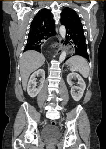
Case Description/Methods: The patient is a 50-year-old male who presents with worsening left upper quadrant pain for the past several months. The pain is described as band-like over the left ribs, constant, tender to palpation, and worse when lying down. Labs were unremarkable. EKG showed sinus bradycardia. CT chest/abdomen/pelvis with contrast showed large hiatal hernia containing a portion of the proximal transverse colon, with no associated bowel obstruction (figure 1).
The patient was diagnosed with an incarcerated Type IV Hiatal Hernia. EGD yielded findings of LA grade D esophagitis and a hiatal hernia. The patient was subsequently scheduled for laparoscopic paraesophageal hernia repair, which yielded a reduced hernia with 3 cm of intra-abdominal esophagus and posterior toupet fundoplication. The patient's symptoms began to resolve, and he was discharged two days later.
Discussion: This case highlights patterns of clinical presentation for Type IV hiatal hernias and its potential to cause bradycardia. The mechanism by which a hiatal hernia causes bradycardia is direct, mechanical compression over the heart or its connected structures. In this case, the large sliding hernia contained a portion of the proximal transverse colon, which likely compromised the vagus nerve, the cardiac chambers, or both, leading to sinus bradycardia.
This case also underscores the importance of a thorough diagnostic approach. Initial workup includes an extensive cardiac enzyme workup, electrocardiogram, and imaging studies. It is only after preliminary differentials in the cardiac workup are determined to be negative that the diagnosis of bradycardia secondary to a Type IV hiatal hernia can be made.
Surgical intervention not only addresses mechanical obstruction, but also alleviates the cardiac symptoms caused by the hernia. Post-surgical improvement in symptoms and resolution of bradycardia further confirms the causative role of the hiatal hernia in this patient’s clinical presentation.

Disclosures:
Sahil Sabharwal, MD, Brandyn Young, BS, Benjamin Boral, DO, Robert Donnell, MD, Kristen Brandon, MD, Sarah Assem, MD. P3763 - A Case of a Type IV Hiatal Hernia Presenting With Sinus Bradycardia, ACG 2024 Annual Scientific Meeting Abstracts. Philadelphia, PA: American College of Gastroenterology.
University of Arkansas for Medical Sciences, Fayetteville, AR
Introduction: A hiatal hernia occurs when contents of the abdomen protrude into the thoracic cavity through the esophageal hiatus. There are four types of hiatal hernias. Type IV hiatal hernias, the most severe, involve the herniation of abdominal viscera, including organs such as the spleen, colon, or small intestine into the thorax. This condition can present with a range of symptoms. However, sinus bradycardia is an especially unique finding of this case report.
Case Description/Methods: The patient is a 50-year-old male who presents with worsening left upper quadrant pain for the past several months. The pain is described as band-like over the left ribs, constant, tender to palpation, and worse when lying down. Labs were unremarkable. EKG showed sinus bradycardia. CT chest/abdomen/pelvis with contrast showed large hiatal hernia containing a portion of the proximal transverse colon, with no associated bowel obstruction (figure 1).
The patient was diagnosed with an incarcerated Type IV Hiatal Hernia. EGD yielded findings of LA grade D esophagitis and a hiatal hernia. The patient was subsequently scheduled for laparoscopic paraesophageal hernia repair, which yielded a reduced hernia with 3 cm of intra-abdominal esophagus and posterior toupet fundoplication. The patient's symptoms began to resolve, and he was discharged two days later.
Discussion: This case highlights patterns of clinical presentation for Type IV hiatal hernias and its potential to cause bradycardia. The mechanism by which a hiatal hernia causes bradycardia is direct, mechanical compression over the heart or its connected structures. In this case, the large sliding hernia contained a portion of the proximal transverse colon, which likely compromised the vagus nerve, the cardiac chambers, or both, leading to sinus bradycardia.
This case also underscores the importance of a thorough diagnostic approach. Initial workup includes an extensive cardiac enzyme workup, electrocardiogram, and imaging studies. It is only after preliminary differentials in the cardiac workup are determined to be negative that the diagnosis of bradycardia secondary to a Type IV hiatal hernia can be made.
Surgical intervention not only addresses mechanical obstruction, but also alleviates the cardiac symptoms caused by the hernia. Post-surgical improvement in symptoms and resolution of bradycardia further confirms the causative role of the hiatal hernia in this patient’s clinical presentation.

Figure: Coronal view of CT chest/abdomen/pelvis showing the type IV hiatal hernia
Disclosures:
Sahil Sabharwal indicated no relevant financial relationships.
Brandyn Young indicated no relevant financial relationships.
Benjamin Boral indicated no relevant financial relationships.
Robert Donnell indicated no relevant financial relationships.
Kristen Brandon indicated no relevant financial relationships.
Sarah Assem indicated no relevant financial relationships.
Sahil Sabharwal, MD, Brandyn Young, BS, Benjamin Boral, DO, Robert Donnell, MD, Kristen Brandon, MD, Sarah Assem, MD. P3763 - A Case of a Type IV Hiatal Hernia Presenting With Sinus Bradycardia, ACG 2024 Annual Scientific Meeting Abstracts. Philadelphia, PA: American College of Gastroenterology.
