Tuesday Poster Session
Category: Colon
P3799 - A Subtle Lesion With an Aggressive Manifestation - A Rare Case of Invasive Squamous Cell Carcinoma of the Hepatic Flexure
Tuesday, October 29, 2024
10:30 AM - 4:00 PM ET
Location: Exhibit Hall E

Has Audio
- JJ
Jason John, DO
Jefferson Health
Pennsauken, NJ
Presenting Author(s)
Jason John, DO1, Anudeep Jala, DO2, Christopher Chhoun, DO3, Joshua Soliman, DO2, Brian Blair, DO3, Lucy Joo, DO3, Richard Walters, DO2, Neethi Dasu, DO4
1Jefferson Health, Pennsauken, NJ; 2Jefferson Health, Washington Township, NJ; 3Jefferson Health, Cherry Hill, NJ; 4Beth Israel Lahey Health, Burlington, MA
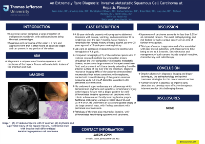
Introduction: Colorectal cancer comprises a large proportion of malignancies worldwide, with adenocarcinoma being the most common type. Squamous cell carcinoma of the colon is a rare and aggressive form that is often found at advanced stages and can present in any portion of the colon. We present a unique case of invasive squamous cell carcinoma of the hepatic flexure with metastatic lesions of the omentum and liver.
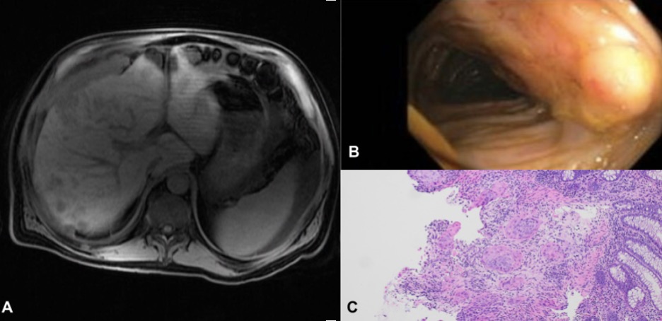
Case Description/Methods: A 56-year-old male presents with progressive abdominal distension with nausea, vomiting, and unintentional 50 lb weight loss over the last 7 months. Social history is remarkable for remote history of heavy alcohol use over 25 years ago and a 20-pack-year smoking history. Lab work on admission revealed macrocytic anemia with hemoglobin of 9.8 g/dL. Computed tomography (CT) of the abdomen/pelvis with IV contrast revealed multiple low-attenuation lesions throughout the liver compatible with hepatic metastatic disease, moderate to large amount of intraperitoneal free fluid, and prominent soft tissue density extending from the anterior surface of the liver into the omentum. Magnetic resonance imaging (MRI) of the abdomen demonstrated innumerable liver lesions consistent with neoplasms, marked soft tissue thickening of the greater omentum measuring up to 4.0 cm AP diameter, consistent with peritoneal carcinomatosis. The patient underwent two large-volume paracentesis with fluid studies demonstrating SAAG < 1.1 and fluid protein >2.5, both for which cytology was negative for malignant cells. Upper endoscopy and colonoscopy demonstrated erythema and superficial inflammatory injury in the hepatic flexure with a biopsy positive for well-differentiated invasive squamous cell carcinoma with colonic submucosa and focally involving lamina propria. Additional malignancy workup revealed CEA of 26 and CA19-9 of 67. He underwent an ultrasound-guided biopsy of the large omental mass, with findings consistent with peritoneal carcinomatosis. Pathology of the mass also returned as invasive, well-differentiated keratinizing squamous cell carcinoma.
Discussion: Squamous cell carcinoma accounts for less than 0.5% of all colorectal cancers. The exact pathophysiology and risk factors for such a unique cancer are an area of further investigation. This type of cancer is aggressive and often associated with poor overall outcomes, with mean survival time being as low as 8.5 months. Early detection and management of such cancers include surgical resection, chemotherapy, and radiotherapy.

Disclosures:
Jason John, DO1, Anudeep Jala, DO2, Christopher Chhoun, DO3, Joshua Soliman, DO2, Brian Blair, DO3, Lucy Joo, DO3, Richard Walters, DO2, Neethi Dasu, DO4. P3799 - A Subtle Lesion With an Aggressive Manifestation - A Rare Case of Invasive Squamous Cell Carcinoma of the Hepatic Flexure, ACG 2024 Annual Scientific Meeting Abstracts. Philadelphia, PA: American College of Gastroenterology.
1Jefferson Health, Pennsauken, NJ; 2Jefferson Health, Washington Township, NJ; 3Jefferson Health, Cherry Hill, NJ; 4Beth Israel Lahey Health, Burlington, MA
Introduction: Colorectal cancer comprises a large proportion of malignancies worldwide, with adenocarcinoma being the most common type. Squamous cell carcinoma of the colon is a rare and aggressive form that is often found at advanced stages and can present in any portion of the colon. We present a unique case of invasive squamous cell carcinoma of the hepatic flexure with metastatic lesions of the omentum and liver.
Case Description/Methods: A 56-year-old male presents with progressive abdominal distension with nausea, vomiting, and unintentional 50 lb weight loss over the last 7 months. Social history is remarkable for remote history of heavy alcohol use over 25 years ago and a 20-pack-year smoking history. Lab work on admission revealed macrocytic anemia with hemoglobin of 9.8 g/dL. Computed tomography (CT) of the abdomen/pelvis with IV contrast revealed multiple low-attenuation lesions throughout the liver compatible with hepatic metastatic disease, moderate to large amount of intraperitoneal free fluid, and prominent soft tissue density extending from the anterior surface of the liver into the omentum. Magnetic resonance imaging (MRI) of the abdomen demonstrated innumerable liver lesions consistent with neoplasms, marked soft tissue thickening of the greater omentum measuring up to 4.0 cm AP diameter, consistent with peritoneal carcinomatosis. The patient underwent two large-volume paracentesis with fluid studies demonstrating SAAG < 1.1 and fluid protein >2.5, both for which cytology was negative for malignant cells. Upper endoscopy and colonoscopy demonstrated erythema and superficial inflammatory injury in the hepatic flexure with a biopsy positive for well-differentiated invasive squamous cell carcinoma with colonic submucosa and focally involving lamina propria. Additional malignancy workup revealed CEA of 26 and CA19-9 of 67. He underwent an ultrasound-guided biopsy of the large omental mass, with findings consistent with peritoneal carcinomatosis. Pathology of the mass also returned as invasive, well-differentiated keratinizing squamous cell carcinoma.
Discussion: Squamous cell carcinoma accounts for less than 0.5% of all colorectal cancers. The exact pathophysiology and risk factors for such a unique cancer are an area of further investigation. This type of cancer is aggressive and often associated with poor overall outcomes, with mean survival time being as low as 8.5 months. Early detection and management of such cancers include surgical resection, chemotherapy, and radiotherapy.

Figure: Image 1: (A) CT abdomen/pelvis with IV contrast, (B) Erythema and superficial injury of the hepatic flexure, (C) Omental mass with invasive well-differentiated keratinizing squamous cell carcinoma
Disclosures:
Jason John indicated no relevant financial relationships.
Anudeep Jala indicated no relevant financial relationships.
Christopher Chhoun indicated no relevant financial relationships.
Joshua Soliman indicated no relevant financial relationships.
Brian Blair indicated no relevant financial relationships.
Lucy Joo indicated no relevant financial relationships.
Richard Walters indicated no relevant financial relationships.
Neethi Dasu indicated no relevant financial relationships.
Jason John, DO1, Anudeep Jala, DO2, Christopher Chhoun, DO3, Joshua Soliman, DO2, Brian Blair, DO3, Lucy Joo, DO3, Richard Walters, DO2, Neethi Dasu, DO4. P3799 - A Subtle Lesion With an Aggressive Manifestation - A Rare Case of Invasive Squamous Cell Carcinoma of the Hepatic Flexure, ACG 2024 Annual Scientific Meeting Abstracts. Philadelphia, PA: American College of Gastroenterology.
