Tuesday Poster Session
Category: Endoscopy Video Forum
P3884 - Single Session Combined Endoscopic and Surgical Treatment of Gallstone Ileus
Tuesday, October 29, 2024
10:30 AM - 4:00 PM ET
Location: Exhibit Hall E

Has Audio

Zehra Naseem, MD
Cleveland Clinic
Cleveland, OH
Presenting Author(s)
Zehra Naseem, MD1, Aun Muhammad, MD2, Muhammad Hamza, 3, Hassan Siddiki, MD4
1Cleveland Clinic, Cleveland, OH; 2University of Mississippi Medical Center, Jackson, MS; 3Aga Khan University, Karachi, Sindh, Pakistan; 4Digestive Disease Institute, Cleveland Clinic, Cleveland, OH
Introduction: Gallstone ileus infrequently causes mechanical bowel obstruction, mostly occurring in the small bowel due to a cholecystoduodenal fistula. Traditionally, managed surgically, there are emerging cases of endoscopic management of gallstones. Here, we describe a unique case where both surgical and endoscopic modalities were employed together for effective management in a single session.
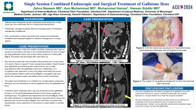
Case Description/Methods: A 61-year-old woman, considered a high-risk surgical candidate due to hypertension, type II diabetes, and stage IV chronic kidney disease, presented to the emergency department with a three-day history of nausea and non-bilious vomiting. A non-contrast abdominal computed tomography scan revealed two large gallbladder stones without evidence of cholecystitis. The patient was subsequently discharged with close follow-up.<br><br>The patient returned a week later with intractable bilious emesis and a five-day history of no stool or flatus. A repeat computed tomography scan revealed pneumobilia in the left hepatic lobe, gallbladder wall thickening with intraluminal air, and a single large gallstone within the gallbladder. Additionally, a 2.5 cm gallstone was observed in a loop of the distal small bowel, causing upstream dilation. A nasogastric tube was placed, and emergent surgery was deferred due to acute renal failure.<br><br>The patient was transferred to our institution for a single-session endoscopic lithotripsy and mini-enterotomy to remove gallstones from the bladder and ileum. A standard upper endoscope with a cap was introduced into the gallbladder through a 10 mm cholecystoduodenal fistula to avoid using a single-use cholangioscope. A 30 mm stone was visualized and fragmented using electrohydraulic lithotripsy, with all fragments removed. While still under anesthesia, a longitudinal enterotomy was performed to remove the culprit 2.5 cm gallstone located 30 cm from the terminal ileum. One month later, the patient was asymptomatic, and an abdominal X-ray showed no bowel dilation or calcified stones.
Discussion: This case highlights the innovative management of gallstone ileus with small bowel obstruction using a combined endoscopic and surgical approach in a single session for a high-risk surgical candidate. We also emphasize the cost-saving benefits achieved by utilizing one anesthesia event and avoiding the use of a single-use direct cholangioscope. Instead, we successfully maneuvered a standard upper scope with a cap to access the gallbladder.
Disclosures:
Zehra Naseem, MD1, Aun Muhammad, MD2, Muhammad Hamza, 3, Hassan Siddiki, MD4. P3884 - Single Session Combined Endoscopic and Surgical Treatment of Gallstone Ileus, ACG 2024 Annual Scientific Meeting Abstracts. Philadelphia, PA: American College of Gastroenterology.
1Cleveland Clinic, Cleveland, OH; 2University of Mississippi Medical Center, Jackson, MS; 3Aga Khan University, Karachi, Sindh, Pakistan; 4Digestive Disease Institute, Cleveland Clinic, Cleveland, OH
Introduction: Gallstone ileus infrequently causes mechanical bowel obstruction, mostly occurring in the small bowel due to a cholecystoduodenal fistula. Traditionally, managed surgically, there are emerging cases of endoscopic management of gallstones. Here, we describe a unique case where both surgical and endoscopic modalities were employed together for effective management in a single session.
Case Description/Methods: A 61-year-old woman, considered a high-risk surgical candidate due to hypertension, type II diabetes, and stage IV chronic kidney disease, presented to the emergency department with a three-day history of nausea and non-bilious vomiting. A non-contrast abdominal computed tomography scan revealed two large gallbladder stones without evidence of cholecystitis. The patient was subsequently discharged with close follow-up.<br><br>The patient returned a week later with intractable bilious emesis and a five-day history of no stool or flatus. A repeat computed tomography scan revealed pneumobilia in the left hepatic lobe, gallbladder wall thickening with intraluminal air, and a single large gallstone within the gallbladder. Additionally, a 2.5 cm gallstone was observed in a loop of the distal small bowel, causing upstream dilation. A nasogastric tube was placed, and emergent surgery was deferred due to acute renal failure.<br><br>The patient was transferred to our institution for a single-session endoscopic lithotripsy and mini-enterotomy to remove gallstones from the bladder and ileum. A standard upper endoscope with a cap was introduced into the gallbladder through a 10 mm cholecystoduodenal fistula to avoid using a single-use cholangioscope. A 30 mm stone was visualized and fragmented using electrohydraulic lithotripsy, with all fragments removed. While still under anesthesia, a longitudinal enterotomy was performed to remove the culprit 2.5 cm gallstone located 30 cm from the terminal ileum. One month later, the patient was asymptomatic, and an abdominal X-ray showed no bowel dilation or calcified stones.
Discussion: This case highlights the innovative management of gallstone ileus with small bowel obstruction using a combined endoscopic and surgical approach in a single session for a high-risk surgical candidate. We also emphasize the cost-saving benefits achieved by utilizing one anesthesia event and avoiding the use of a single-use direct cholangioscope. Instead, we successfully maneuvered a standard upper scope with a cap to access the gallbladder.
Disclosures:
Zehra Naseem indicated no relevant financial relationships.
Aun Muhammad indicated no relevant financial relationships.
Muhammad Hamza indicated no relevant financial relationships.
Hassan Siddiki: Boston Scientific consulting – Consultant.
Zehra Naseem, MD1, Aun Muhammad, MD2, Muhammad Hamza, 3, Hassan Siddiki, MD4. P3884 - Single Session Combined Endoscopic and Surgical Treatment of Gallstone Ileus, ACG 2024 Annual Scientific Meeting Abstracts. Philadelphia, PA: American College of Gastroenterology.

