Tuesday Poster Session
Category: Colon
P3771 - Blastoid Mantle Cell Lymphoma Presenting as a Cecal Mass
Tuesday, October 29, 2024
10:30 AM - 4:00 PM ET
Location: Exhibit Hall E

Has Audio

Lefika Bathobakae, MD
St. Joseph's University Medical Center
Paterson, NJ
Presenting Author(s)
Lefika Bathobakae, MD1, Jorge Lopez, MD2, Rammy Bashir, MD3, Heba Farhan, BS4, Mohamed Elagami, MD2, Kamal Amer, MD5, Yana Cavanagh, MD5
1St. Joseph's University Medical Center, Paterson, NJ; 2St. Joseph’s University Medical Center, Paterson, NJ; 3St. George’s University School of Medicine, Grenada, West Indies, Paterson, NJ; 4Rowan-Virtua University School of Osteopathic Medicine, Paterson, NJ; 5St. Joseph's University Medical Center, Paterson, NJ
Introduction: Blastoid mantle cell lymphoma (BV-MCL) is a rare and aggressive subtype of B-cell non-Hodgkin’s lymphoma that can spread to the GI tract. Endoscopic features can range from normal-appearing mucosa to multiple lymphomatous polyposis (MLP). MCL presenting as an isolated cecal mass is exceedingly rare and a diagnostic and therapeutic challenge. Herein, we present a rare case of BV-MCL presenting as an isolated cecal mass.
Case Description/Methods: A 64-year-old male with a history of HTN and a pancreatic head lesion (1.4 cm) presented to the ED complaining of acute-onset diffuse abdominal pain and melena. He denied hematemesis, acid reflux, acute dysphagia, diarrhea, or hematochezia. On exam, the abdomen was soft, diffusely tender on light palpation, but non-peritoneal. Digital rectal exam showed black tarry stools without palpable masses, fissures, or perianal ulcers. Triage labs: WBC, 11.8x10³/mm3 and Hgb, 10.8 g/dL.
Contrast-enhanced CT scan of the chest, abdomen and pelvis revealed a large cecal mass measuring 6.9 x 6.0 cm, with regional and peripancreatic/porta hepatis lymphadenopathy. Tumor markers were unremarkable. MRCP showed a cecal mass with extensive lymphadenopathy.
EGD with EUS showed diffuse pancreatic lobularity and normal main pancreatic duct. Gastric biopsies showed moderate chronic inactive gastritis. Colonoscopy revealed nodular mucosa in the ascending colon and in the cecum. An ulcerated and partially obstructing large mass was found in the cecum (Figure 1). Histopathology revealed mantle cell lymphoma with blastoid morphology and a high proliferation index. On immunostaining, the atypical lymphoid cells were diffusely positive for CD20, CD5, cyclin D1, Bcl-2 and CD43. The B cells were negative for CD10, BCL6, MUM1 and TdT. The patient's hospital course was complicated by mechanical bowel obstruction due to cecal mass. The obstruction resolved after one cycle of Bendamustine/rituximab chemotherapy. So far, the patient has completed two cycles of chemotherapy of the anticipated four and is fully active.
Discussion: MCL is frequently seen in the form of MLP in which innumerable polyps are observed in the GI tract. In rare instances, MCL can present as a cecal mass. Signs and symptoms of MCL in the GI tract include abdominal pain, bleeding, and intestinal obstruction. Treatment options include chemotherapy regimens such as R-CHOP (rituximab, cyclophosphamide, doxorubicin, vincristine, and prednisone) and RB (Rituximab and Bendamustine).

Disclosures:
Lefika Bathobakae, MD1, Jorge Lopez, MD2, Rammy Bashir, MD3, Heba Farhan, BS4, Mohamed Elagami, MD2, Kamal Amer, MD5, Yana Cavanagh, MD5. P3771 - Blastoid Mantle Cell Lymphoma Presenting as a Cecal Mass, ACG 2024 Annual Scientific Meeting Abstracts. Philadelphia, PA: American College of Gastroenterology.
1St. Joseph's University Medical Center, Paterson, NJ; 2St. Joseph’s University Medical Center, Paterson, NJ; 3St. George’s University School of Medicine, Grenada, West Indies, Paterson, NJ; 4Rowan-Virtua University School of Osteopathic Medicine, Paterson, NJ; 5St. Joseph's University Medical Center, Paterson, NJ
Introduction: Blastoid mantle cell lymphoma (BV-MCL) is a rare and aggressive subtype of B-cell non-Hodgkin’s lymphoma that can spread to the GI tract. Endoscopic features can range from normal-appearing mucosa to multiple lymphomatous polyposis (MLP). MCL presenting as an isolated cecal mass is exceedingly rare and a diagnostic and therapeutic challenge. Herein, we present a rare case of BV-MCL presenting as an isolated cecal mass.
Case Description/Methods: A 64-year-old male with a history of HTN and a pancreatic head lesion (1.4 cm) presented to the ED complaining of acute-onset diffuse abdominal pain and melena. He denied hematemesis, acid reflux, acute dysphagia, diarrhea, or hematochezia. On exam, the abdomen was soft, diffusely tender on light palpation, but non-peritoneal. Digital rectal exam showed black tarry stools without palpable masses, fissures, or perianal ulcers. Triage labs: WBC, 11.8x10³/mm3 and Hgb, 10.8 g/dL.
Contrast-enhanced CT scan of the chest, abdomen and pelvis revealed a large cecal mass measuring 6.9 x 6.0 cm, with regional and peripancreatic/porta hepatis lymphadenopathy. Tumor markers were unremarkable. MRCP showed a cecal mass with extensive lymphadenopathy.
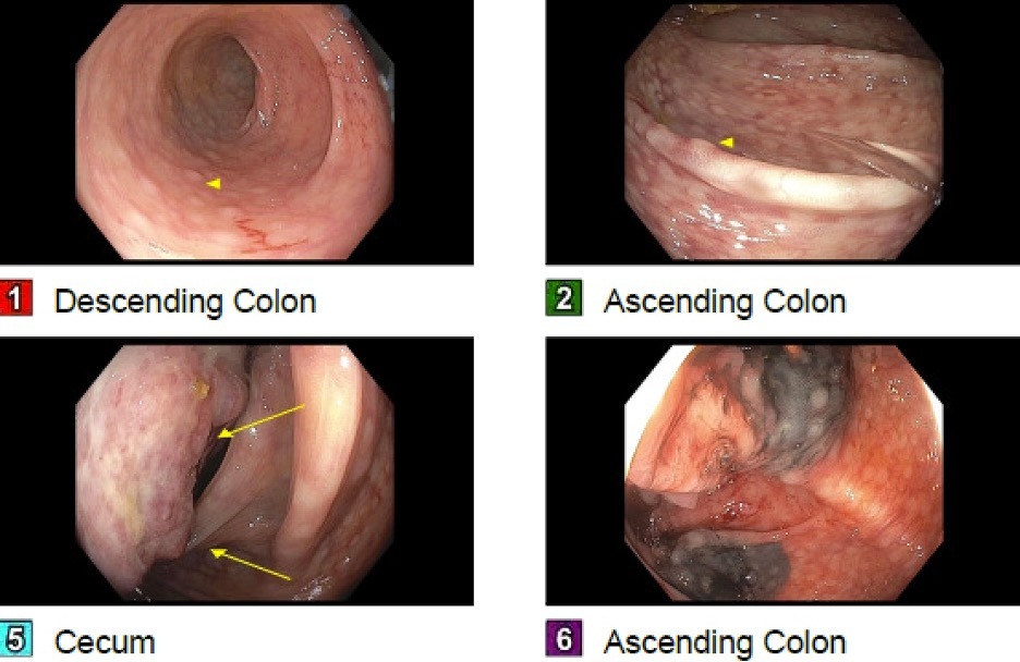
EGD with EUS showed diffuse pancreatic lobularity and normal main pancreatic duct. Gastric biopsies showed moderate chronic inactive gastritis. Colonoscopy revealed nodular mucosa in the ascending colon and in the cecum. An ulcerated and partially obstructing large mass was found in the cecum (Figure 1). Histopathology revealed mantle cell lymphoma with blastoid morphology and a high proliferation index. On immunostaining, the atypical lymphoid cells were diffusely positive for CD20, CD5, cyclin D1, Bcl-2 and CD43. The B cells were negative for CD10, BCL6, MUM1 and TdT. The patient's hospital course was complicated by mechanical bowel obstruction due to cecal mass. The obstruction resolved after one cycle of Bendamustine/rituximab chemotherapy. So far, the patient has completed two cycles of chemotherapy of the anticipated four and is fully active.
Discussion: MCL is frequently seen in the form of MLP in which innumerable polyps are observed in the GI tract. In rare instances, MCL can present as a cecal mass. Signs and symptoms of MCL in the GI tract include abdominal pain, bleeding, and intestinal obstruction. Treatment options include chemotherapy regimens such as R-CHOP (rituximab, cyclophosphamide, doxorubicin, vincristine, and prednisone) and RB (Rituximab and Bendamustine).

Figure: Figure 1. Endoscopic image showing an ulcerated partially obstructing large mass in the cecum and nodular mucosa in the ascending colon.
Disclosures:
Lefika Bathobakae indicated no relevant financial relationships.
Jorge Lopez indicated no relevant financial relationships.
Rammy Bashir indicated no relevant financial relationships.
Heba Farhan indicated no relevant financial relationships.
Mohamed Elagami indicated no relevant financial relationships.
Kamal Amer indicated no relevant financial relationships.
Yana Cavanagh indicated no relevant financial relationships.
Lefika Bathobakae, MD1, Jorge Lopez, MD2, Rammy Bashir, MD3, Heba Farhan, BS4, Mohamed Elagami, MD2, Kamal Amer, MD5, Yana Cavanagh, MD5. P3771 - Blastoid Mantle Cell Lymphoma Presenting as a Cecal Mass, ACG 2024 Annual Scientific Meeting Abstracts. Philadelphia, PA: American College of Gastroenterology.
